|
|
 |
|
|
|
|
|
A 24-year-old contact lens wearer with unilateral vision loss requiring penetrating keratoplasty
Digital Journal of Ophthalmology 2019
Volume 25, Number 2
June 30, 2019
DOI: 10.5693/djo.03.2019.06.001
|
Printer Friendly
Download PDF |
|
|
 Jonathan T. L. Lee, MBBS, BMedSc
 | Department of Ophthalmology, Alfred Health, Melbourne, Victoria, Australia Chengde Pham, MBBS, BMedSci | Department of Ophthalmology, Alfred Health, Melbourne, Victoria, Australia Edward Greenrod, MBBS, FRANZCO | Department of Ophthalmology, Alfred Health, Melbourne, Victoria, Australia
|
|
|
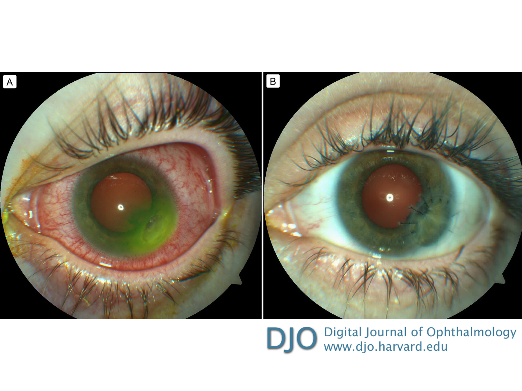
| Examination | | On initial examination, best-corrected visual acuity was 20/16 in the right eye and 20/80 in the left eye. Pupils were equal and reactive, with no afferent pupillary defect. Slit-lamp examination of the left eye revealed a dense 2.4 × 3.7 mm corneal infiltrate, with a surrounding 8.0 mm immune ring (Figure 1A). There was 2+ anterior chamber cell and flare, with associated ciliary injection. Posterior segment examination showed no vitreous cells and healthy fundus. Intraocular pressure was 9 mm Hg in the affected left eye. The fellow eye was unremarkable. | |
|
Figure 1
A, Dense corneal infiltrate with conjunctival injection at presentation. B, Clear corneal graft 9 weeks after penetrating keratoplasty. Best-corrected visual acuity was 20/40.
 |
|
|
|
|
|
|
|
 Welcome, please sign in