|
|
 |
|
|
|
|
|
A 45-year-old man with unilateral optic disc edema and vision loss
Digital Journal of Ophthalmology 2019
Volume 25, Number 1
March 29, 2019
DOI: 10.5693/djo.03.2019.02.002
|
Printer Friendly
Download PDF |
|
|
 Benjamin G. Jastrzembski, MD
 | Division of Ophthalmology, Beth Israel Deaconess Medical Center, Harvard Medical School, Boston, Massachusetts; Department of Ophthalmology, Hospital for Sick Children, University of Toronto, Toronto, Ontario Nurhan Torun, MD | Division of Ophthalmology, Beth Israel Deaconess Medical Center, Harvard Medical School, Boston, Massachusetts
|
|
|
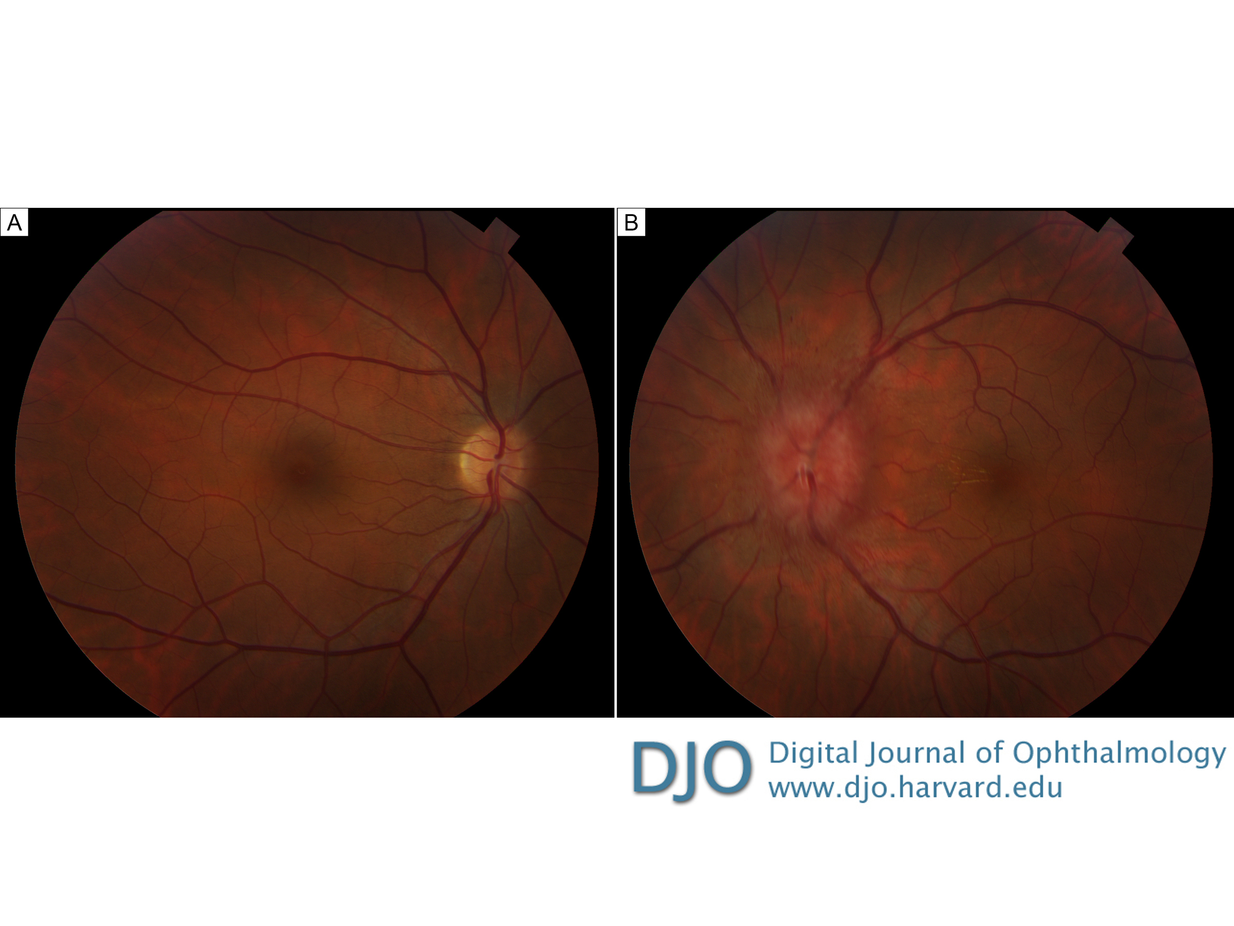
| Examination | | On examination, visual acuity was 20/20 in the right eye and 20/50 in the left eye, with a relative afferent pupillary defect in the left eye. Extraocular motility was full. The patient identified 8/8 Ishihara color plates in the right eye and 0/8 color plates in the left eye. The anterior segment examination was unremarkable in both eyes. On dilated fundus examination, there was trace vitreous cell and diffuse disc edema in the left eye, with dilation of the peripapillary vessels; there was a partial macular star in the left eye. The right eye appeared normal (Figure 1). | |
|
Figure 1
Initial fundus photographs demonstrating a normal posterior pole in the right eye and diffuse disc edema, dilation of the peripapillary vessels, and macular star in the left eye.
 |
|
|
|
|
|
|
|
 Welcome, please sign in