|
|
 |
|
|
|
|
|
A 45-year-old man with unilateral optic disc edema and vision loss
Digital Journal of Ophthalmology 2019
Volume 25, Number 1
March 29, 2019
DOI: 10.5693/djo.03.2019.02.002
|
Printer Friendly
Download PDF |
|
|
 Benjamin G. Jastrzembski, MD
 | Division of Ophthalmology, Beth Israel Deaconess Medical Center, Harvard Medical School, Boston, Massachusetts; Department of Ophthalmology, Hospital for Sick Children, University of Toronto, Toronto, Ontario Nurhan Torun, MD | Division of Ophthalmology, Beth Israel Deaconess Medical Center, Harvard Medical School, Boston, Massachusetts
|
|
|
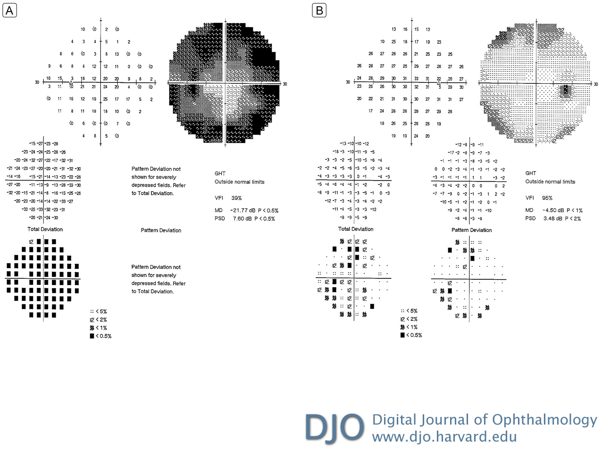
| Ancillary Testing | Humphrey 30-2 SITA-Fast automated visual field testing demonstrated scattered nonspecific loss in the right eye and generalized dense depression in the left eye, with a mean deviation of −4.50 dB in the right eye and −21.77 dB in the left eye (Figure 2). The following laboratory studies were drawn and results were unremarkable: complete blood count with differential, Bartonella hensalae IgG and IgM antibodies, Lyme disease IgG and IgM antibodies, Toxoplasma gondii IgG and IgM antibodies, fluorescent treponemal antibody absorption (FTA-ABS), rapid plasma reagin (RPR), neuromyelitis optica IgG antibody (Aquaporin 4 protein antibody), angiotension converting enzyme (ACE) level, anti-neutrophil cytoplasmic antibody (ANCA), IgG subclasses, and serum protein electrophoresis.
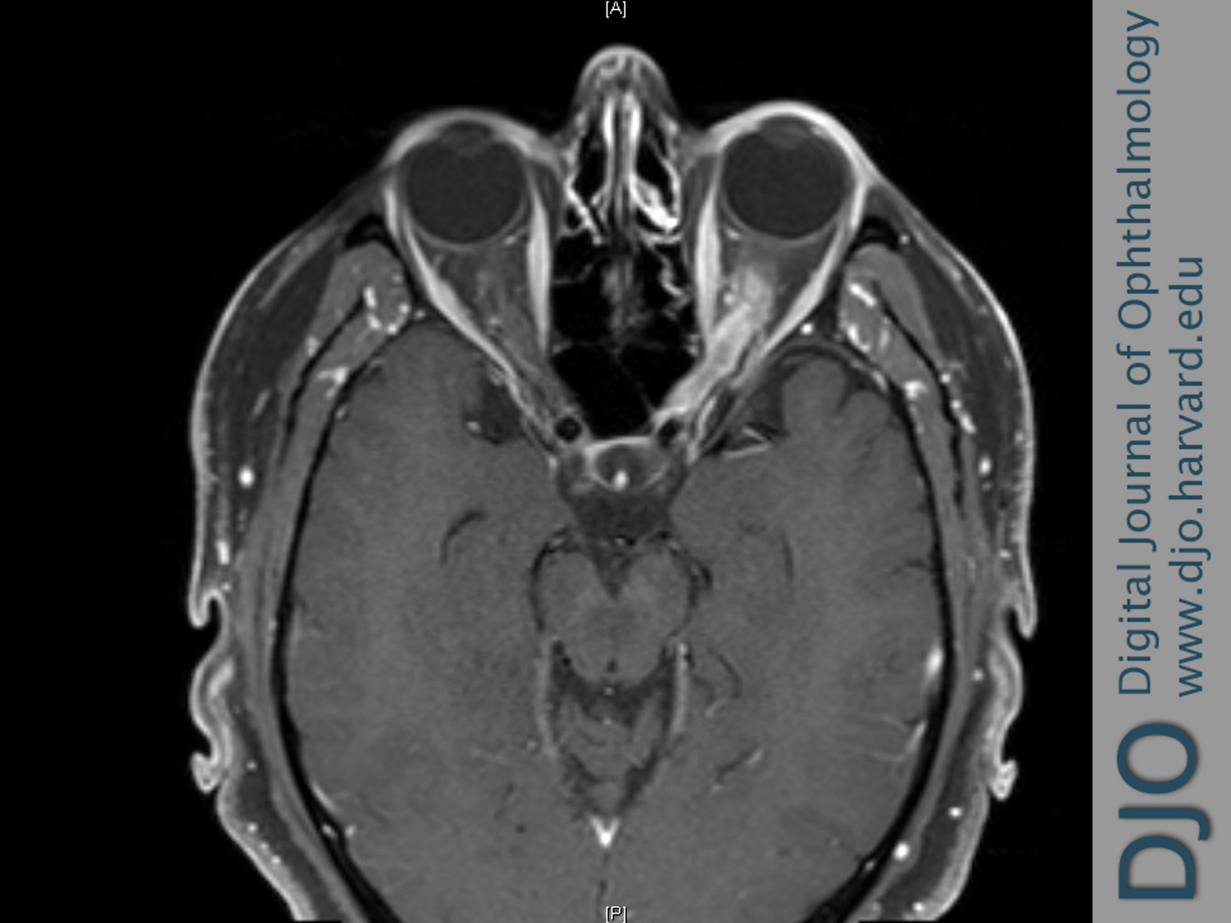
Magnetic resonance imaging (MRI) of the brain with and without contrast was obtained and revealed tram track enhancement of the left optic nerve sheath, with no white matter lesions or other abnormalities (Figure 3).
Additional studies were performed, including a lumbar puncture, which revealed normal cerebrospinal fluid constituents, negative cytology, and insufficient cells to perform flow cytometry. Whole-body PET computed tomography was also negative for signs of sarcoidosis or malignancy. | |
|
Figure 2
Initial Humphrey 30-2 SITA-fast automated visual field testing with scattered nonspecific loss in the right eye (B) and generalized depression with relative central sparing in the left eye (A).
 |
|
|
Figure 3
T1-weighted, post-contrast axial magnetic resonance imaging (MRI) of the brain demonstrating tram track enhancement of the left optic nerve sheath complex.
 |
|
|
|
|
|
|
|
 Welcome, please sign in