Vision:20/20 OU uncorrected
Color Plates:Normal color vision OU
Pupils: Equal, reactive, No APD
Amsler grid: no metamorphopsia OU
Visual Field: Confrontational visual fields demonstrated a large temporal scotoma in the right eye; the left eye was full to confrontation.
Motility: Full OU
Applanation pressure: 19 mmHg OD, 13 mm Hg OS
Slit lamp examination:normal anterior segments OU
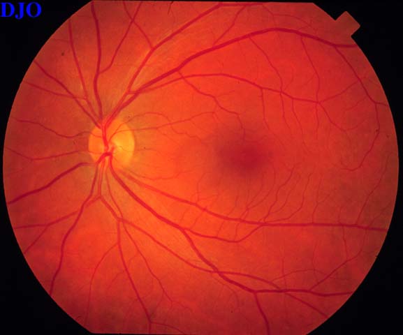
Fundus examination: See Figures 1-3
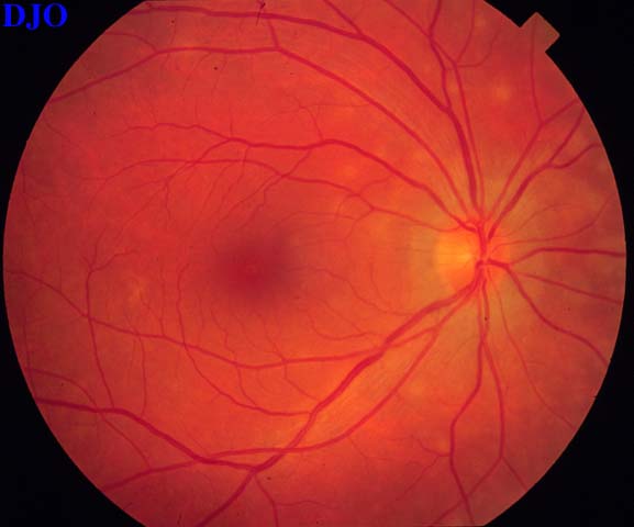
Figure 1
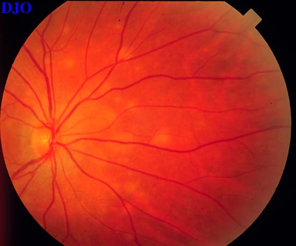
Figures 1-2. Ophthalmoscopy of the right eye revealed small, discrete white lesions at the level of the deep retina or retinal pigment epithelium scattered over the posterior pole. More lesions are noted nasally. The optic disc and macula were normal.
 Welcome, please sign in