|
|
 |
|
|
|
|
|
22 year old female comes in for a normal eye exam
Digital Journal of Ophthalmology 1997
Volume 3, Number 27
October 21, 1997
|
Printer Friendly
|
|
|
|
|
|
|
| Ancillary Testing | Angiogram
| |
|
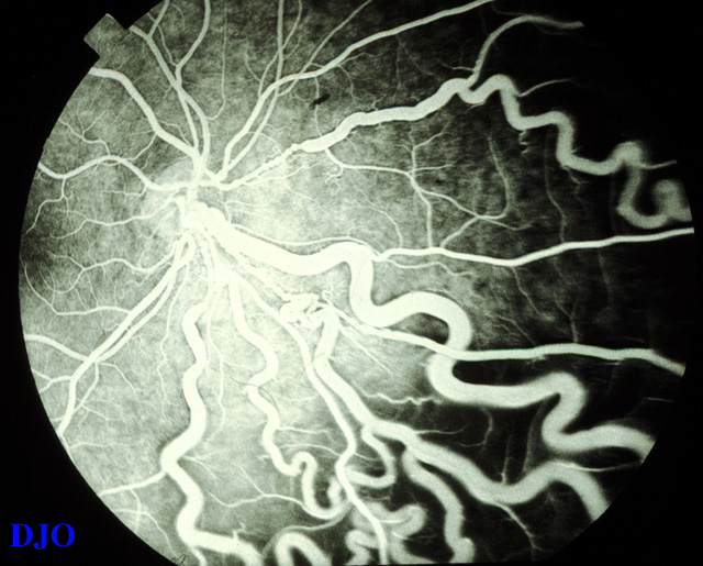
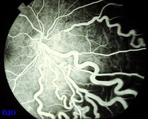
Figure 3
(OS) Fluorescein angiogram of the large dilated vessels.
 |
|
|
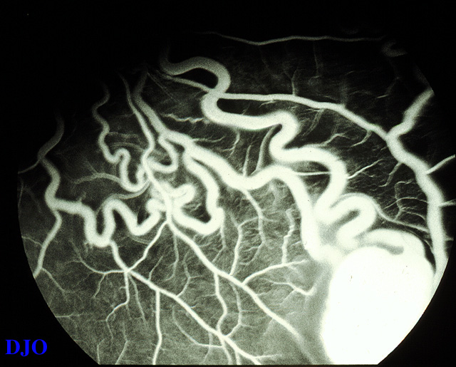
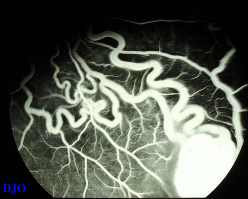
Figure 4
(OS) Fluorescein angiogram of the vascular tumor. It hyperfluoresces in the early phases and the feeder vessels fill. Aside FROM the feeder vessels, the surrounding vasculature appears normal.
 |
|
|
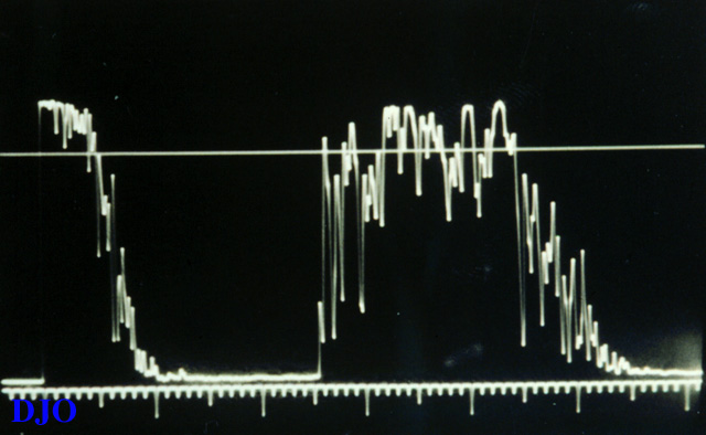
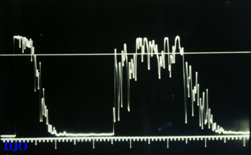
Figure 5
(OS) A-scan ultrasound of the lesion shows a 2.3 mm elevated mass with a medium to high reflectivity and a slightly irregular internal structure.
 |
|
|
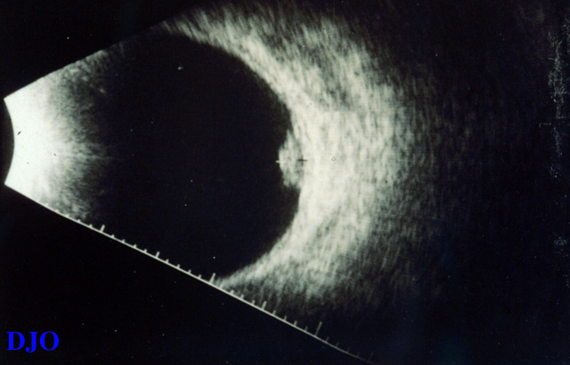
Figure 6
(OS) B-scan ultrasound of the lesion shows a slightly irregular highly reflective dome shaped lesion in the inferotemporal fundus extending to the equator.
 |
|
|
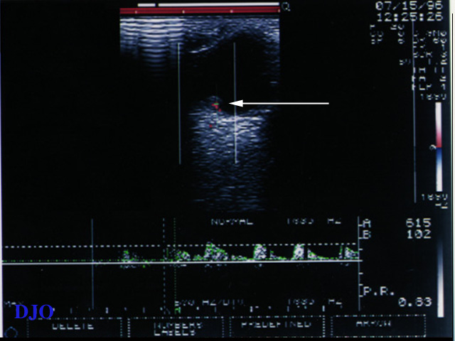
Figure 7
(OS) Color Doppler of the lesion (white arrow) shows minimal high flow vascularity in the lesion or the feeder vessels.
 |
|
|
|
|
|
 Welcome, please sign in