|
|
 |
|
|
|
|
|
25 year old kickboxer with progressive proptosis
Digital Journal of Ophthalmology 1997
Volume 3, Number 26
September 24, 1997
|
Printer Friendly
|
|
|

Scott C. Brun, MD | Massachusetts Eye and Ear Infirmary, Harvard Medical School, Boston, MA Peter A.D. Rubin, M.D. | Massachusetts Eye and Ear Infirmary, Harvard Medical School, Boston, MA
|
|
|
| Ancillary Testing | Radiographic Studies
Pathology
| |
|
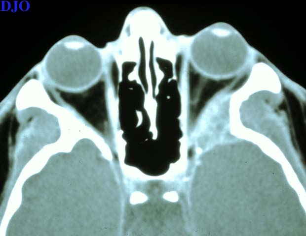
Figure 2a
Orbital CT: Axial, showing an expansile soft tissue mass obliterating the greater wing of the sphenoid bone.
 |
|
|
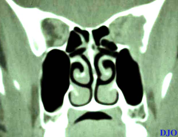
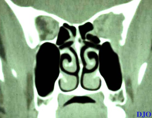
Figure 2b
Orbital CT: Coronal bone window, showing scalloping of the greater wing of the sphenoid bone indicating bony destruction.
 |
|
|
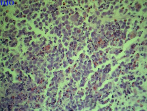
Figure 3
Histology revealed a number of large histiocytes with scattered eosinophills, lymphocytes and plasma cells.
 |
|
|
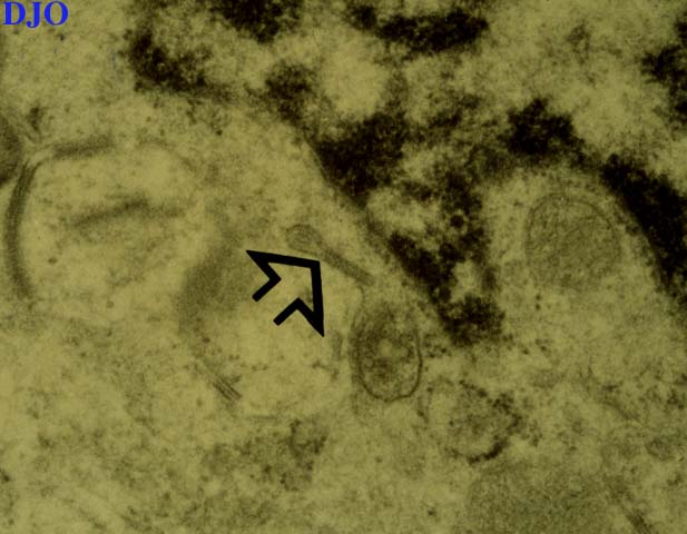
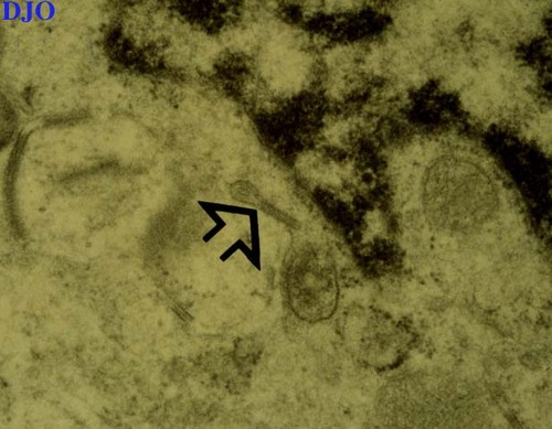
Figure 4
Electron microscopy demonstrated the presence of Birbeck granules (arrow) within a few of the histiocytes.
 |
|
|
|
|
|
 Welcome, please sign in