|
|
 |
|
|
|
|
|
A 69 year old woman with a painful, red, right eye for two days
Digital Journal of Ophthalmology 1997
Volume 3, Number 19
May 28, 1997
|
Printer Friendly
|
|
|

Brian P. Connolly, MD | Wills Eye Hospital, Philadelphia, PA Marlon Maus, MD | Wills Eye Hospital, Philadelphia, PA
|
|
|
| Examination | Vision: 20/20 OD 20/25 OS sc
Pupils: Equal, briskly reactive to light. No RAPD
Slit lamp examination: Normal OU
Extraocular Movements: Normal OU
Fundus examination: Normal OU
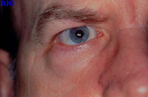
External: A firm, cystic mass was affixed to the lateral aspect of the orbital floor just posterior to the orbital rim. The posterior border of this lesion could not be appreciated clinically. | |
|
Figure 1
This is a photograph of the patient's right eye and orbit
 |
|
|
|
|
|
 Welcome, please sign in


