|
|
 |
|
|
|
|
|
A 69 year old woman with a painful, red, right eye for two days
Digital Journal of Ophthalmology 1997
Volume 3, Number 19
May 28, 1997
|
Printer Friendly
|
|
|

Brian P. Connolly, MD | Wills Eye Hospital, Philadelphia, PA Marlon Maus, MD | Wills Eye Hospital, Philadelphia, PA
|
|
|
| Ancillary Testing | Radiographic Studies
Pathology
| |
|
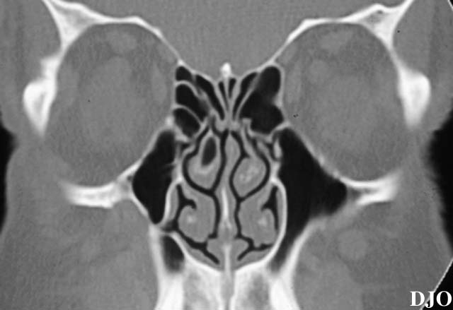
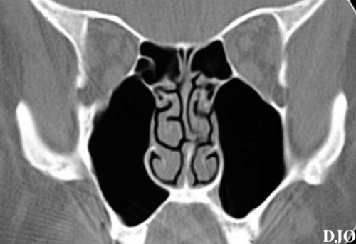
Figure 2
Figures 2-3. A non-contrast CT of the orbits (coronal images) was obtained. A lesion corresponding to the clinical mass was noted in the inferolateral aspect of the right orbit affixed to the orbital bone. The mass was only slightly more radio-dense than the surrounding orbital fatty tissues, and its margins were not clearly demarcated. There were several areas where the orbital bones overlying the maxillary sinus appeared to be eroded by this mass.
 |
|
|
Figure 3
 |
|
|
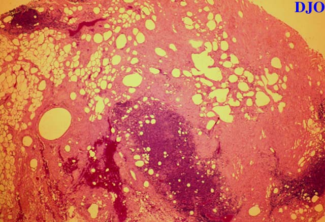
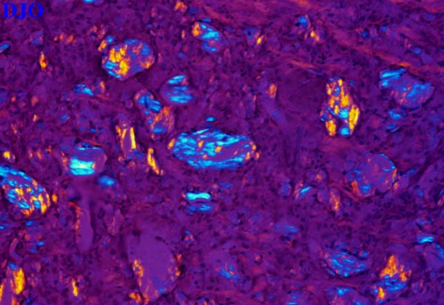
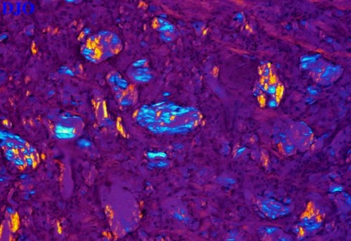
Figure 4
Figures 4-5. Pathology Specimens An orbitotomy with excisional biopsy was performed. Pathologic examination of frozen sections disclosed fibrosis and chronic granulomatous inflammation centered around foci of birefringent foreign material (Figure 4.). The birefringent material was noted to dissolve in processing and paraffin embedding suggesting a lipid composition (Figure 5.). These findings were consistent with a diagnosis of paraffinoma with foreign body inflammatory reaction.
 |
|
|
Figure 5
 |
|
|
|
|
|
|
|
 Welcome, please sign in