Vision: 16/40--> 16/15 (PH) OD; 16/200 OS Pupils: 2+ left afferent pupillary defect External exam: Normal Motility: Full, slight increase in left periorbital pain with extreme right gaze Slit lamp examination: Normal ou. Anterior chambers deep and quiet. Intraocular pressure: 15 mm Hg OU Visual field Examination: See Figures 1-2 Fundus examination: See Figure 3-4
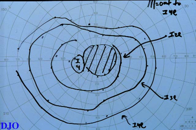
Figure 1
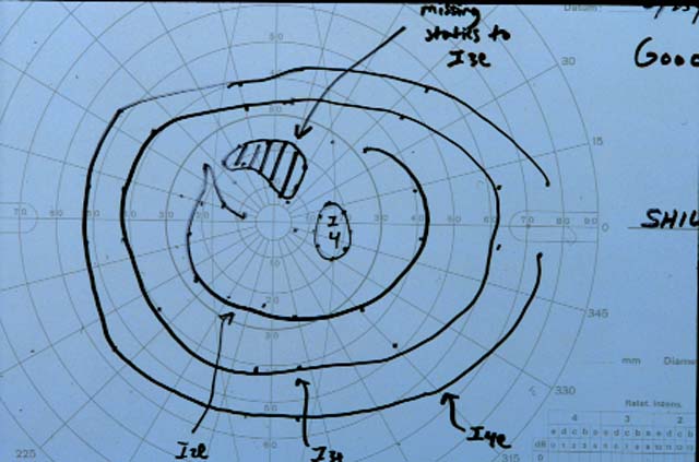
Figures 1-2. Goldmann visual fields are shown. There is a dense central scotoma OS. In addition, there was a paracentral visual field defect OD.
Figure 2
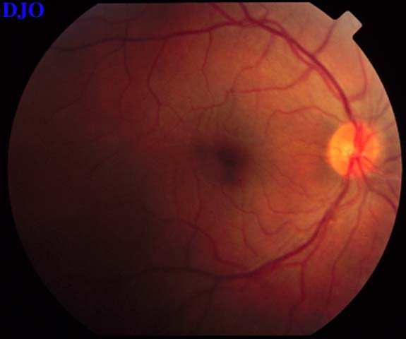
Figure 3
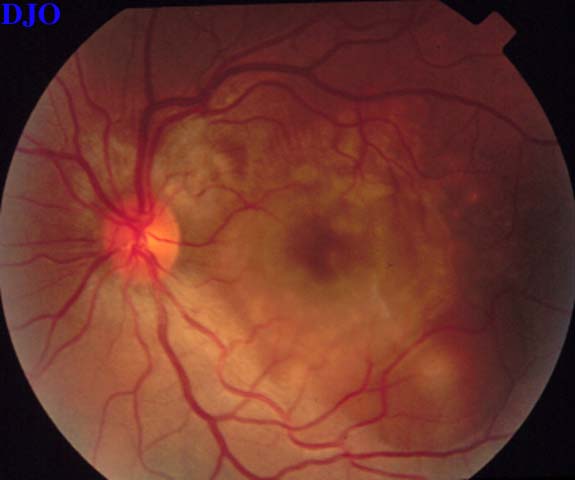
Figures 3-4. The right and left fundi are shown. The right fundus demonstrated fine striae in the macula, which were evident with slitlamp biomicroscopy. The left fundus showed an elevated serous retinal detachment of the macula and the peripapillary retina. The left disk appeared slightly hyperemic, but appeared otherwise normal.
 Welcome, please sign in