|
|
 |
|
|
|
|
|
3 year old boy with swelling under the left eye
Digital Journal of Ophthalmology 1997
Volume 3, Number 17
April 16, 1997
|
Printer Friendly
|
|
|

Mina Massaro, MD | Scheie Eye Institute, University of Pennsylvania Medical School, Philadelphia, PA James A Katowitz MD | Scheie Eye Institute, University of Pennsylvania Medical School, Philadelphia, PA
|
|
|
| Diagnosis and Discussion | Retained Wooden Foreign Body
Clinical Course:
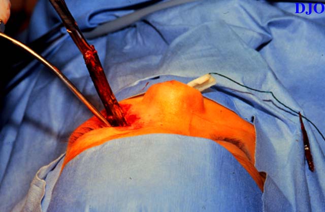
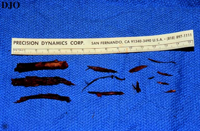
The patient was admitted and started on IV antibiotics and brought to the OR for removal of the foreign body. Surgery was done under nasal endoscopic visualization and pieces of bark and a 5 cm branch was removed FROM the nasal cavity through the original wound site.
Wooden foreign bodies can appear as a lucency similar to air or fat on CT scan and plain X-ray. If a wooden object is suspected, consider obtaining an MRI which will image wood better. Wood matter in the orbit or brain can harbor soil organisms which can lead to panophthalmitis, meningitis, and even death. It is imperative that retained wooden foreign bodies be diagnosed and treated immediately.
Because children are unable to provide detailed histories, one should always be suspicious of serious injuries in the setting of trauma even if the initial clinical examination is unremarkable. | |
|
Figure 2
Figures 2-3. On the left, the length of the branch can be seen as it is surgically removed. On the right, fragments of the branch which were removed are shown in comparison with a 15 cm ruler
 |
|
|
Figure 3
 |
|
|
|
|
|
|
|
 Welcome, please sign in