|
|
 |
|
|
|
|
|
3 year old boy with swelling under the left eye
Digital Journal of Ophthalmology 1997
Volume 3, Number 17
April 16, 1997
|
Printer Friendly
|
|
|

Mina Massaro, MD | Scheie Eye Institute, University of Pennsylvania Medical School, Philadelphia, PA James A Katowitz MD | Scheie Eye Institute, University of Pennsylvania Medical School, Philadelphia, PA
|
|
|
| Ancillary Testing | Radiographic Studies
| |
|
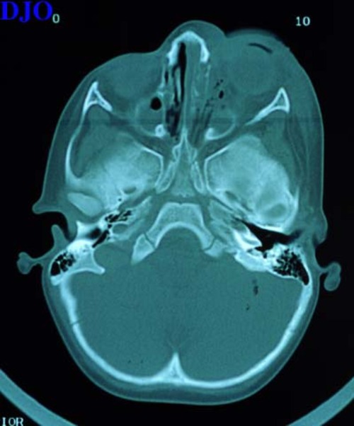
Figure 1
As was noted in the history, initial CT scanning revealed a left medial wall fracture. Repeat CT scan of orbits revealed penetrating trauma by a cylindrical foreign body imbedded in the nasal cavity which caused fractures to the left medial orbit wall, nasal septum, and lateral nasal wall.
 |
|
|
|
|
|
 Welcome, please sign in


