|
|
 |
|
|
|
|
|
A 15-year-old-boy with an optic neuropathy
Digital Journal of Ophthalmology 2017
Volume 23, Number 4
December 5, 2017
DOI: 10.5693/djo.03.2015.06.001
|
Printer Friendly
Download PDF |
|
|

David B. Lazar, MD | Department of Ophthalmology, Louisiana State University, New Orleans, Louisiana Adham B. Hariri, MD | Department of Ophthalmology, Ochsner Clinic Foundation, New Orleans, Louisiana
|
|
|
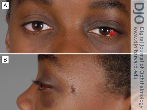
| Examination | | On clinical examination, the patient had mild edema and ecchymosis of the left upper eyelid, mild proptosis of the left eye, a small subconjunctival hemorrhage in the temporal conjunctiva, and commotio retinae in his left macula. Ishihara color plates were full in both eyes. There was no resistance to retropulsion and no relative afferent pupillary defect. He was given oral antibiotics and a three-day course of oral steroids. He was followed closely and 1 week later referred to the oculoplastic surgery service for evaluation of the orbital foreign body. At the time of this evaluation (Figure 1), his visual acuity in the left eye had decreased to 20/40, and he had developed a mild left afferent pupillary defect. In addition, there was restriction of gaze to the extreme right secondary to pain. | |
|
Figure 1
External photographs taken 7 days following the incident showing the temporal subconjunctival hemorrhage, eyelid ecchymosis, and mild proptosis in the left eye (A) and the entry site of the pellet in the left temple (B).
 |
|
|
|
|
|
|
|
 Welcome, please sign in


