|
|
 |
|
|
|
|
|
A 15-year-old-boy with an optic neuropathy
Digital Journal of Ophthalmology 2017
Volume 23, Number 4
December 5, 2017
DOI: 10.5693/djo.03.2015.06.001
|
Printer Friendly
Download PDF |
|
|

David B. Lazar, MD | Department of Ophthalmology, Louisiana State University, New Orleans, Louisiana Adham B. Hariri, MD | Department of Ophthalmology, Ochsner Clinic Foundation, New Orleans, Louisiana
|
|
|
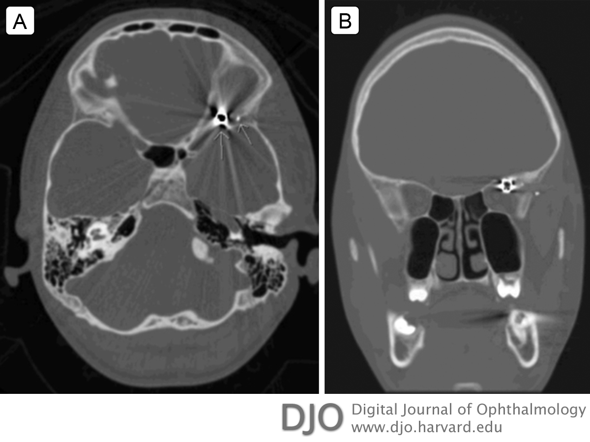
| Ancillary Testing | | A computed tomography (CT) scan showed a 4.5 mm metallic foreign body within the posterior and superior aspect of the left orbital apex in close proximity to the optic nerve. A second 1-2 mm foreign body fragment was located adjacent to the superficial aspect of the lateral orbital wall along the inner aspect of the left temporalis muscle (Figure 2A). Visualization of the lateral rectus muscle was limited secondary to imaging artifact of the adjacent foreign body. A defect was noted in the left lateral orbital wall with a comminuted fracture and osteoid formation, indicating that the foreign body had entered through the lateral wall (Figure 2B). The globe, calvarium, calvarial soft tissues, and brain were noted to be unremarkable. | |
|
Figure 2
Computed tomography of the head. A, Axial scan showing the 4.5 mm metallic foreign body located in the apex of the left orbit; also visualized is a smaller shrapnel fragment lodged in the inner aspect of the left temporalis muscle. B, Coronal scan showing the path of the pellet through the left temporalis muscle and fractured lateral orbital wall, stopping short of and immediately adjacent to the optic nerve within the apex.
 |
|
|
|
|
|
|
|
 Welcome, please sign in