|
|
 |
|
|
|
|
|
19 year old man with a "corneal abrasion"
Digital Journal of Ophthalmology 1997
Volume 3, Number 15
March 18, 1997
|
Printer Friendly
|
|
|

Yichieh Shiuey, MD | Massachusetts Eye and Ear Infirmary, Harvard Medical School, Boston, MA Kathy Colby, MD | Massachusetts Eye and Ear Infirmary, Harvard Medical School, Boston, MA Claes Dohlman, MD | Massachusetts Eye and Ear Infirmary, Harvard Medical School, Boston, MA
|
|
|
| Examination | Vision: 20/70 OD; 20/30 OS
Pupils: Reactive OU, No relative afferent pupillary defect
External exam: Mild ptosis OU with fissure heights of 6 mm in each eye
Slit lamp examination: See Figures 1-5
Fundus examination: Within normal limits OU
Management: The patient was started on pred forte q2hrs OD, cromolyn qid OU, cyclogyl bid OD, ciloxan BID OD, bandage CL OD. Corneal cultures were taken and the patient was referred to an allergist. | |
|
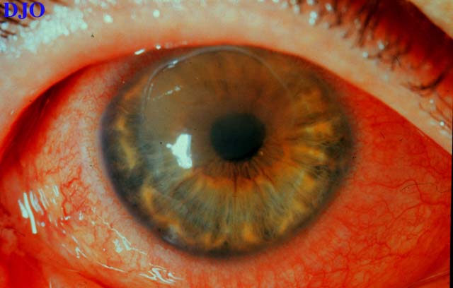
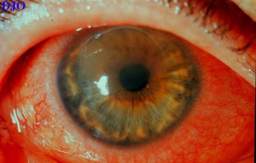
Figure 1
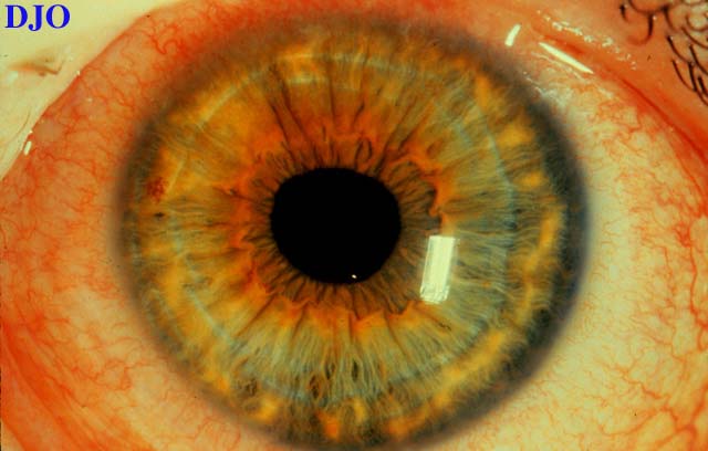
Figures 1-2. In the right eye, there was 3+ conjunctival injection, a superior epithelial defect, microcystic edema, and superficial corneal haze. In the left eye, there was 1+ conjunctival injection
 |
|
|
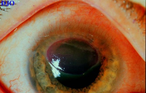
Figure 2
 |
|
|
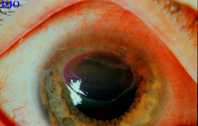
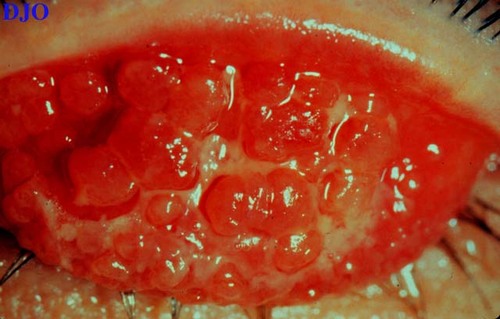
Figure 3
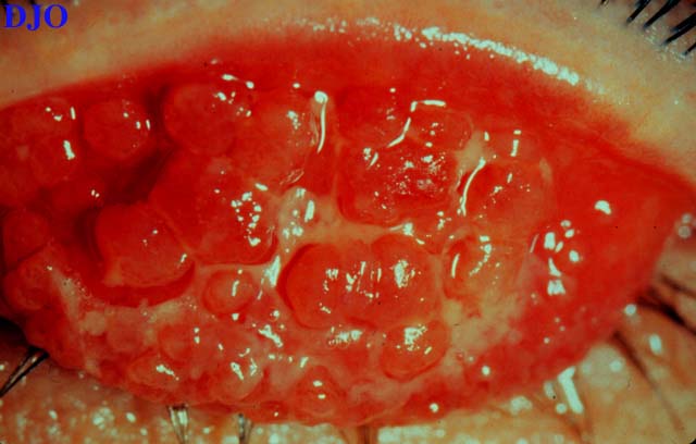
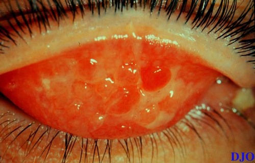
Figures 3-5. The epithelial defect on the right cornea was well dilineated with rose bengal stain. Lid eversion revealed large cobblestone pappilae OU which were covered in a tenacious mucous.
 |
|
|
Figure 4
 |
|
|
Figure 5
 |
|
|
|
|
|
|
|
 Welcome, please sign in