|
|
 |
|
|
|
|
|
39 year old Hispanic man who presents with decreased vision OS for one year
Digital Journal of Ophthalmology 1996
Volume 2, Number 10
December 24, 1996
|
Printer Friendly
|
|
|

John A. Irvine, M.D. | Doheny Eye Institute, University of Southern California, Los Angeles, California Irene C. Kuo, M.D. | Doheny Eye Institute, University of Southern California, Los Angeles, California
|
|
|
| Examination | Vision: 20/25 OD, 20/50 OS
Pupils: No APD
External: leonine facies
Slit Lamp Exam:
- Lids: Absence of lashes OU. Loss of the tarsal plate more marked in the upper lids than in the lower ones OU; lid margins thickened OU
- Conjunctiva: Grade one plus follicles OU
- Sclera: quiet OU
- Cornea: punctate epithelial opacities superiorly OU with pannus formation.; prominent anterior stromal opacities; decreased corneal sensation in all quadrants.
- Anterior chamber: deep and quiet OU
Tonometry: Normal OU
Schirmer's Test: Normal OU
Fundus examination: The disc, macula, and vessels OU were within normal limits. | |
|
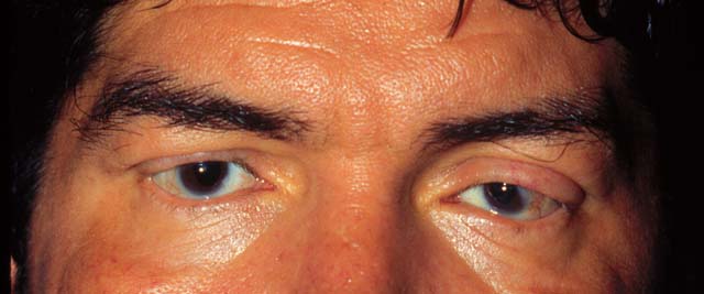
Figure 1
This is a racoon view of the patient's eyes. Please note the loss of eyelashes.
 |
|
|
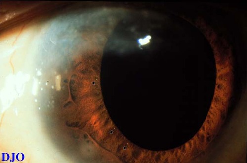
Figure 2
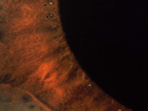
Figures 2-3. These are low and high power views of the patient's anterior segments. There are small discrete lesions on the surface of the iris, which are pointed out by the black arrows.
 |
|
|
Figure 3
 |
|
|
|
|
|
|
|
 Welcome, please sign in