Vision: CF at 4 ft OD ph 20/200; 20/30 OS ph 20/20 Pupils: Normal OU, No APD Slit lamp examination: See Figure 1 Applanation Tonometry: 15 mmHG OU Fundus examination: Normal OU Corneal Topography: See Figure 2
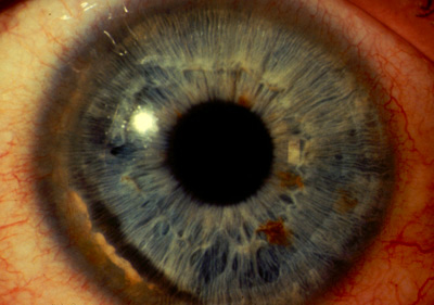
Figure 1
Slit-lamp photo of the right eye showing an area of 90% thinning peripherally, extending FROM the 6:00 to the 1:00 o'clock position. Neovascular changes within the furrow are present with an intact epithelium and a leading edge of lipid deposition anteriorly. The left eye showed similar but much milder changes with a 10% thinning over a three clock hour area.
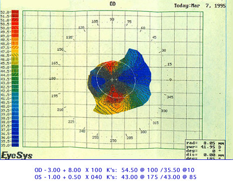
Figure 2
This is the patient's manifest refraction, K readings, and corneal topography.
 Welcome, please sign in