J Oscar Croxatto, MD | Fundacion Oftalmologica Argentina Jorge Malbran Ricardo Negroni, MD | Hospital Municipal F. J. Muiz Gabriela Volpe, MD | Hospital Oftalmologico Santa Luca
Pathology
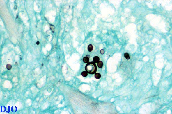
A biopsy of the right eyelid showed an ulcerated skin lesion with pseudoepitheliomatous epidermal hyperplasia. The tissue was infiltrated by a mixture of suppurative and granulomatous inflammation with multinucleated giant cells containing round structures. Grocott stain revealed the presence of intracellular and extracellular budding round yeasts, some of them with a ship's-wheel configuration consistent with Paracoccidioides brasiliensis.
Figure 2
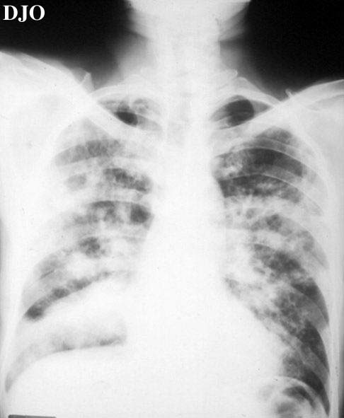
A chest radiograph showed multiple bilateral nodular lesions of the lung with collapse of the inferior right lobe. Computed tomography revealed a cavitated lesion with an air-fluid level.
 Welcome, please sign in