|
|
 |
|
|
|
|
|
39 year old asymptomatic woman with a history of "glaucoma"
Digital Journal of Ophthalmology 1996
Volume 2, Number 6
October 25, 1996
|
Printer Friendly
|
|
|
|
|
|
|
| Examination | Vision: 20/20 OD, 20/20 OS
Pupils: No APD
External: Normal OU
Slit lamp examination: The conjunctiva and sclera were white and quiet OU. The corneas were clear OU. The anterior chamber had no cell or flare.
Tonometry: 15 mmHG OD, 38 mmHG OS
Fundus examination: The left disc shows evidence of glaucomatous cupping with a thin temporal rim. The disc, macula, and vessels OD were within normal limits.
Visual Fields: Testing using the Humphrey automated perimeter showed glaucomatous field loss OS. The visual field was normal OD. | |
|
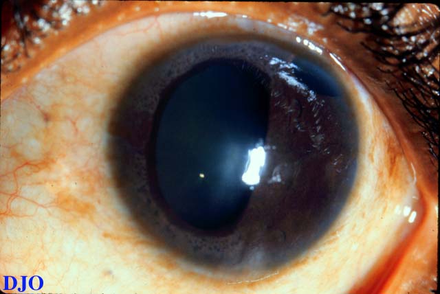
Figure 1
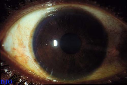
Figures 1-2. These are low magnification photographs of the patient's anterior segments which SHOW distortion of the patient's left pupil (without dilation). The patient's right anterior segment is normal.
 |
|
|
Figure 2
 |
|
|
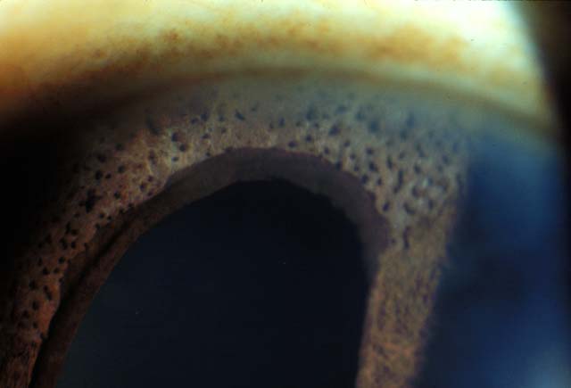
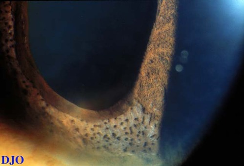
Figure 3
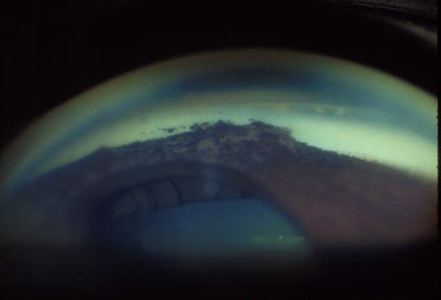
Figures 3-5. Figure 3 and 4 demonstrate multiple fine iris nodules in the patient's left eye. Figure 5 is a gonio-view demonstrating a large area of peripheral anterior synechiae which pulls on the iris and distorts the pupil.
 |
|
|
Figure 4
 |
|
|
Figure 5
 |
|
|
|
|
|
|
|
 Welcome, please sign in