|
|
 |
|
|
|
|
|
48 year old man with bilateral, progressive visual loss over 20 years
Digital Journal of Ophthalmology 1996
Volume 2, Number 5
October 22, 1996
|
Printer Friendly
|
|
|

Dale Heuer, MD | Doheny Eye Institute, University of Southern California, Los Angeles, California Randy Nguyen, MD | Doheny Eye Institute, University of Southern California, Los Angeles, California Michael B. Yang, MD | Doheny Eye Institute, University of Southern California, Los Angeles, California
|
|
|
| Examination | Vision: 20/30 OD, 20/25 OS
Pupils: Normal OU, No APD
Motility: Full OU
Color: Normal OU
External: Normal OU
Slit lamp examination: Normal OU
Tonometry: 16 OU, ranges FROM 14-18 OU during diurnal curve measurements
Gonioscopy: Grade 4 angles, open 360 degrees without pigment, iris processes, synechiae, or bombe OU
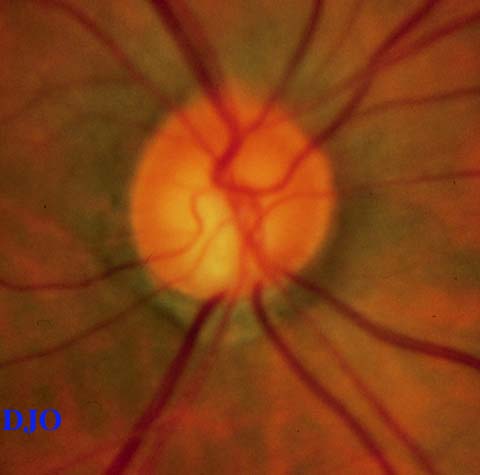
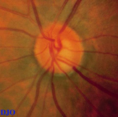
Fundus examination: Macula and vessels appeared normal, the disks are shown in Figures 1-2
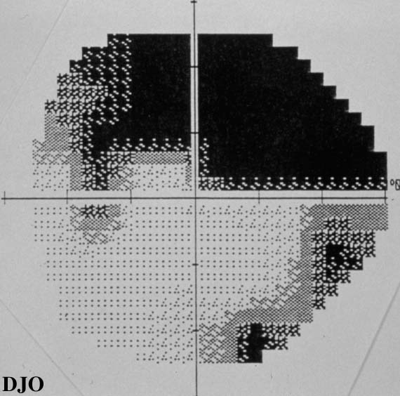
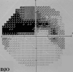
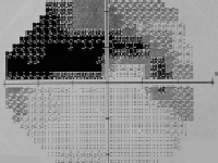
Visual fields: See Figures 3-4 | |
|
Figure 1
Figures 1-2. Disks SHOW cupping and inferior temporal notching OU
 |
|
|
Figure 2
 |
|
|
Figure 3
Figures 3-4. Arcuate defects present in both visual fields.
 |
|
|
Figure 4
 |
|
|
|
|
|
|
|
 Welcome, please sign in