Vision: 20/200 OD, 20/20 OS Pupils: Normal OU, No APD External: Slight scarring on the face and trunk in previous areas of rash Slit lamp examination: Normal OU, anterior chamber and anterior vitreous clear Fundus examination: see Figures 1-3
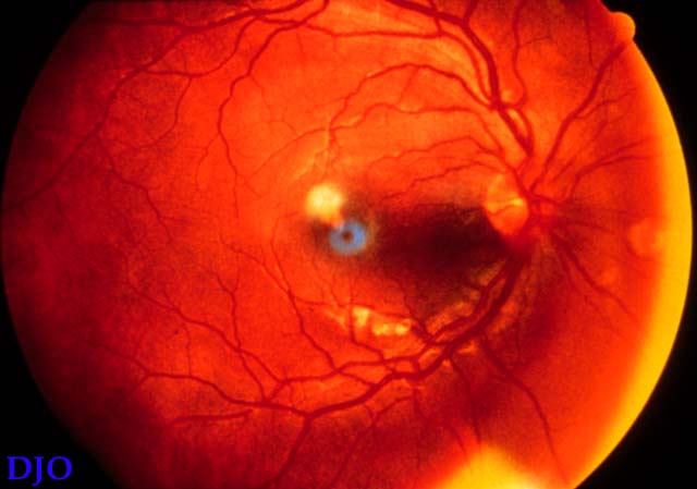
Figure 1
This is a view of the right fundus at presentation, there is a whitish perifoveal lesion in the macula
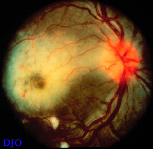
Figure 2
View of the right fundus, one week after presentation which shows extension of the whitish lesion, the lesion appears to involve the deep retina with relative sparing of the retinal vessels.
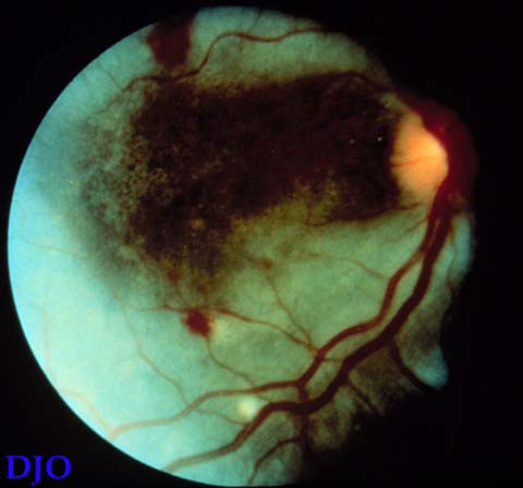
Figure 3
View of the right fundus at four weeks which shows a complete wipe out of the posterior pole by the lesion, there is also a large amount of hemorrhage present. At this point the patient had NLP vision.
 Welcome, please sign in