|
|
 |
|
|
|
|
|
26 year old female with decreased visual acuity
Digital Journal of Ophthalmology 1999
Volume 5, Number 10
April 8, 1999
|
Printer Friendly
|
|
|

Melissa Torralva, MD | Stanford University School of Medicine; Stanford, California 94305 Saad Shaikh, MD | Stanford University School of Medicine; Stanford, California 94305
|
|
|
| Examination | Vision: 20/300 (OD), 20/300 (OS)
Pupils: Normal (OU), no APD
Motility: Full (OU)
Tonometry: Normal (OU)
External: Normal (OU)
Slit Lamp Examination: Normal (OU)
Fundus Exam: Figures 1-4
Upon presentation three weeks later the patientís visual acuity had improved to 20/25 (OU) and fundus examination revealed that most of the hemorrhages had resolved. The patientís hemoglobin and platelet count had also steadily improved. She was still however being treated for active leukemic disease. | |
|
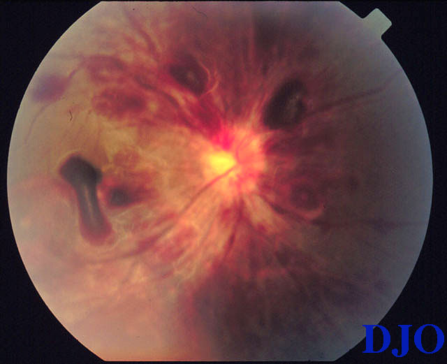
Figure 1
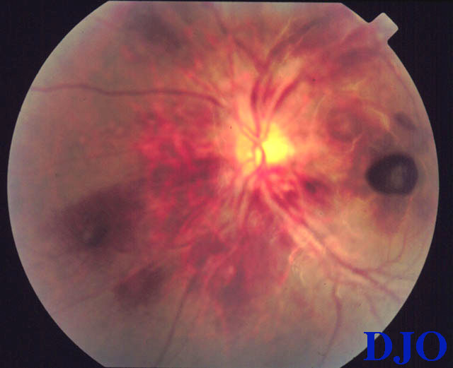
Figures 1-2. Preretinal, intraretinal, and subretinal hemorrhages along with some areas of sub-RPE hemorrhages are present. Mild retinal edema is also present
 |
|
|
Figure 2
 |
|
|
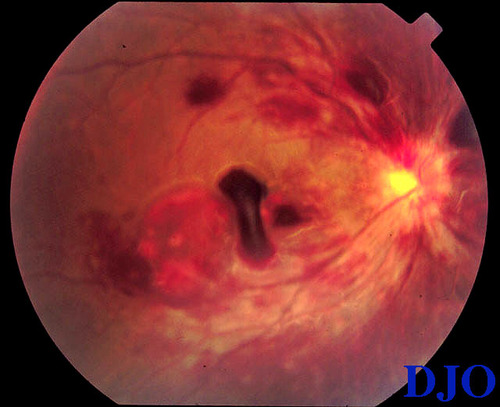
Figure 3
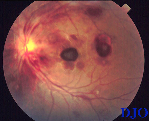
Figures 3-4. Peripapillary never fiber layer and intraretinal hemorrhages are present but the optic nerve is otherwise normal.
 |
|
|
Figure 4
 |
|
|
|
|
|
|
|
 Welcome, please sign in