|
|
 |
|
|
|
|
|
4 year old boy with proptosis of the left eye and an afferent pupillary defect
Digital Journal of Ophthalmology 1999
Volume 5, Number 9
February 8, 1999
|
Printer Friendly
|
|
|

Peter A.D. Rubin, M.D.,F.A.C.S. | Massachusetts Eye and Ear Infirmary, Harvard Medical School, Boston, MA Ramin Tayani, M.D., M.P.H. | Massachusetts Eye and Ear Infirmary, Harvard Medical School, Boston, MA
|
|
|
| Treatment | Surgical Management
The diagnosis of lymphangioma with an acute hemorrhage was highest in our differential. Given the presence of an orbital comparment syndrome (elevated intraorbital pressure resulting INTO an optic nerve compromise) we elected to perform an urgent drainage and biopsy of the mass. The lesion was approached via a lateral orbitotomy following an emergent canthtomy and cantholysis. The mass was found in the intraorbital space, DISTINCT FROM the orbital fat and extraocular muscles. Upon incision of the cystic structure copious amount of venous-appearing blood drained FROM the lesion. Immediately and dramatically the orbit was decongested.
Post-operative report
On post-operative day 1, CT was repeated and an axial image is shown as in Figure 4.
The patient had reversal of his pre-operative afferent pupillary defect, proptosis, extraocular motility limitation. His vision of the right eye was back to 20/25. A focus of nasal chemosis vs. lymphangiectasis had not changed. | |
|
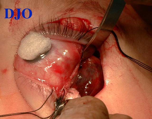
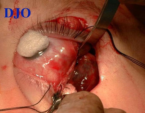
Figure 4
Intra-operative photograph showing the "chocolate cyst" caused by the hemorrhage INTO the lesion.
 |
|
|
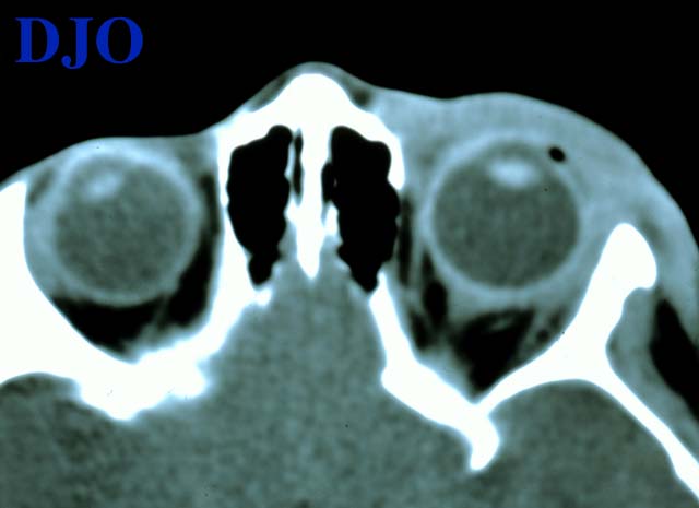
Figure 5
Post-operative (day 1) Axial CT (non-contrast): Significant relief of proptosis, absence of orbital mass, and resolution of globe contour irregularity. Soft tissue changes (edema) in the pre-septal area persisted.
 |
|
|
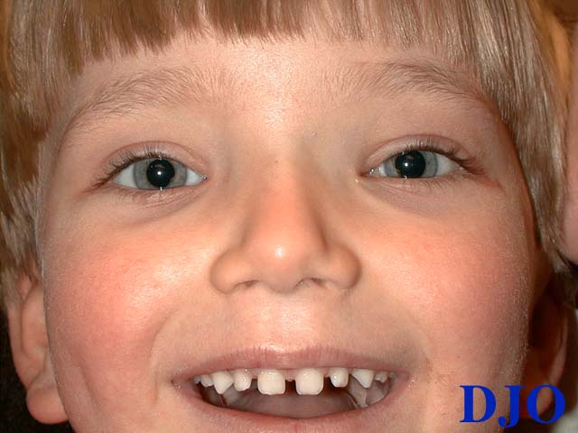
Figure 6
Four months post-operative photo
 |
|
|
|
|
|
|
|
 Welcome, please sign in