Peter A.D. Rubin, M.D.,F.A.C.S. | Massachusetts Eye and Ear Infirmary, Harvard Medical School, Boston, MA Ramin Tayani, M.D., M.P.H. | Massachusetts Eye and Ear Infirmary, Harvard Medical School, Boston, MA
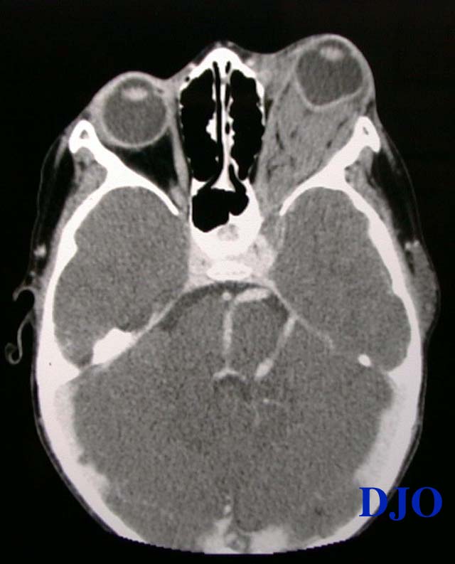
Figure 2
Orbital CT with contrast: Axial: An irregularly-shaped retrobulbar orbital mass filling the posterior orbit with near obliteration of the orbital fat. The severe proptosis and mass effect of the lesion have resulted in an abnormal globe contour. The lesion appears to extend posteriorly through the superior orbital fissure and there is enlargement of the ipsilateral cavernous sinus. A large tortous ipsilateral vascular anomaly is noted. The adjacent paranasal sinuses are clear.
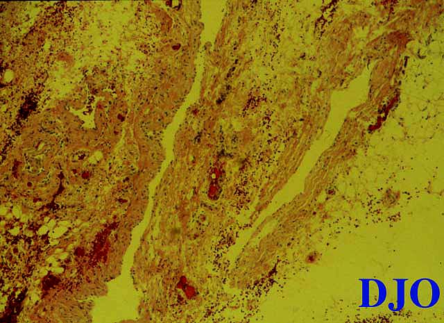
Figure 3
The histopathology is composed of lymph-filled spaces of variable sizes. The spaces are lined by attenuated endothelium characteristic of lymphatic channels.
 Welcome, please sign in