|
|
 |
|
|
|
|
|
29 year old woman with loss of visual field in her right eye
Digital Journal of Ophthalmology 1999
Volume 5, Number 8
January 8, 1999
|
Printer Friendly
|
|
|
|
|
|
|
| Examination | Vision: CF OD improved to 20/20 with raised chin; OS 20/20
Confrontational visual fields: Superior Visual Field restriction, OD; full OS
Pupils: 4 --> 2 OD 4 --> 2 OS
Motility: Full OS
Slit lamp examination: Conjunctiva- white/quite/ no chemosis OU Cornea- clear/quiet OU Anterior chamber- deep/quiet OU Iris- normal, round pupil OU Lens- clear OU
Tonometry: 17 mmHg, OU
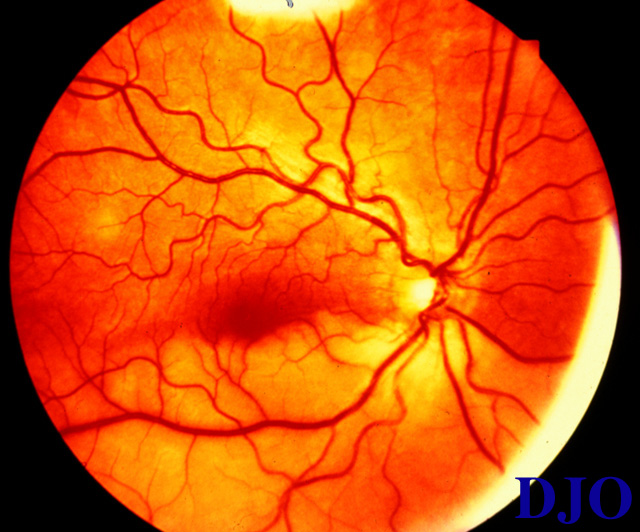
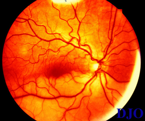
Fundi: On dilated funduscopic examination of the right, diffuse whitening of inferior retina of the right eye extending FROM 3 O'clock to 9 O'clock with retinal edema with arteriolar attenuation (Figure 1). No embolus was visible in any retinal arterioles. The left eye fundus was normal.
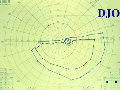
Goldmanns' Field Test: see Figure 2
FA: Fluorescein angiography was deferred by patient
Hematologic work-up:
"0">
|
Full Blood Count |
All parameters normal |
ESR : |
11mm/ 1 hour |
Auto-antibody screen |
Normal |
Routine Coagulation |
Normal |
Anti Thrombin III |
84 units (75-150) |
Total protein S |
60 units (80-115) |
Protein C |
68 units (70-140) |
Blood Chemistry |
Normal |
Echo Cardiography: Normal
Doppler Carotid Angiogram: Normal | |
|
Figure 1
Right eye fundus photo on presentation dedmonstrating inferior retinal whitening and edema with arteriolar attenuation
 |
|
|
Figure 2
Goldmanns' Visual Field testing demonstrating sharply demarcated superior visual field loss in the right eye
 |
|
|
|
 Welcome, please sign in