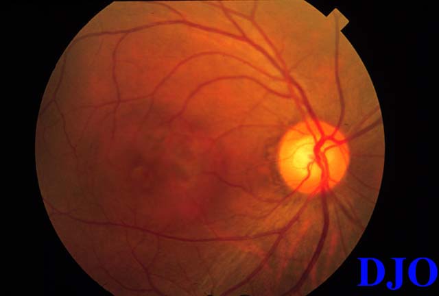
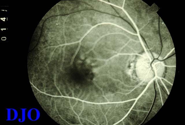
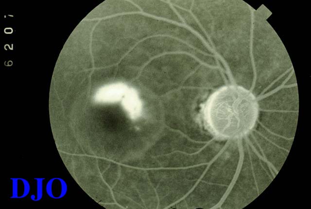
Vision: OD 20/60 distance & near (no improvement with pinhole); OS 20/25 distance & near Confrontational visual fields: full Pupils: 4 --> 2 OD 4 --> 2 OS Motility: Full OS Slit lamp examination: Conjunctiva- white/quite/ no chemosis OU Cornea- clear/quiet OU Anterior chamber- deep/quiet OU Iris- normal, round pupil OU Lens- clear OU Tonometry: OD 22, OS 21 Fundi: On dilated funduscopic examination of the right, a shallow detachment of the macula was seen, with serous subretinal fluid. The optic disk, vitreous, and periphery were normal. Funduscopic examination of the left eye was unremarkable. FA: Fluorescein angiography was performed. The right eye had multiple RPE window defects and leak points in the perifoveal area.
 Welcome, please sign in