Welcome, please
sign in
Original Articles
Case Reports
Grand Rounds
Images
Editorials
Reviews
MEEI Grand Rounds Videos
Archive >
Knowledge Review
Patient Info
Grand Rounds
Most Recent Cases
Dates of Case
Type of Case
Submit a
Grand Round
.
Register
with DJO to receive personalized updates.
If you're already a
member, please
sign in
.
68 year old man with left orbital fullness
Digital Journal of Ophthalmology 1998
Volume 4, Number 23
October 6, 1998
Printer Friendly
Ammar Safar, M.D. | Georgetown University, Washington D.C.
HISTORY
EXAMINATION
ANCILLARY TESTING
DIFF. DIAGNOSIS
DIAGNOSIS AND DISCUSSION
Examination
Vision:
20/20 OD, 20/25 OS
Pupils:
Normal OU
Motility:
Mild restriction to adduction OS
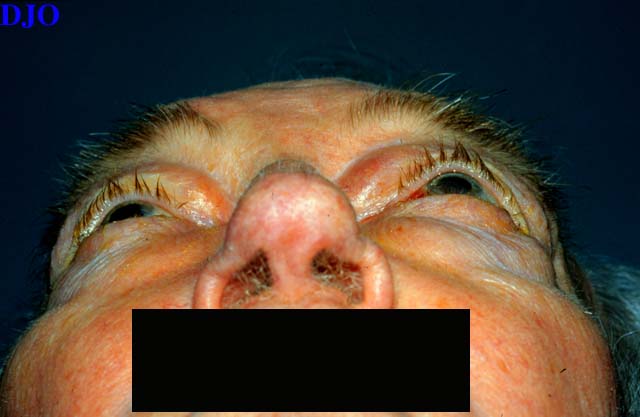
External exam:
see Figures 1 and 2
Slit lamp examination:
Normal OU
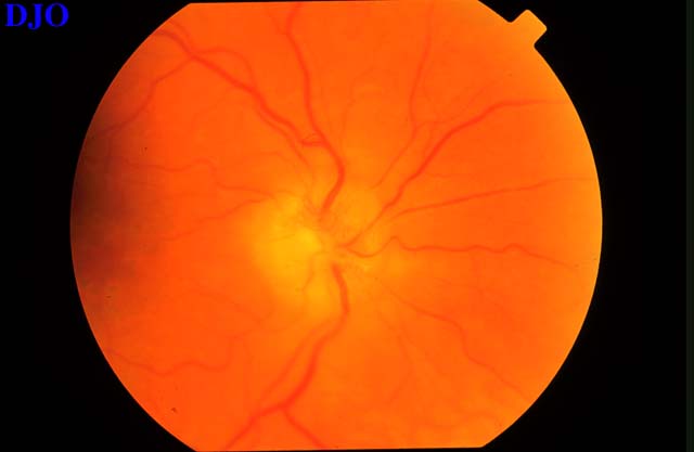
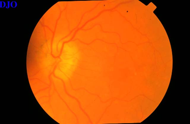
Fundus examination:
see Figures 3 and 4
top
Figure 1
Figures 1-2. Mild proptosis OS with left superonasal orbital mass, which was firm and non-mobile on palpation
Figure 2
Figure 3
Figures 3-4. Chronic disk swelling OU. Macula and periphery normal OU.
Figure 4
 Welcome, please sign in