|
|
 |
|
|
|
|
|
68 year old man with left orbital fullness
Digital Journal of Ophthalmology 1998
Volume 4, Number 23
October 6, 1998
|
Printer Friendly
|
|
|
|
|
|
|
| Ancillary Testing | Radiographic Studies
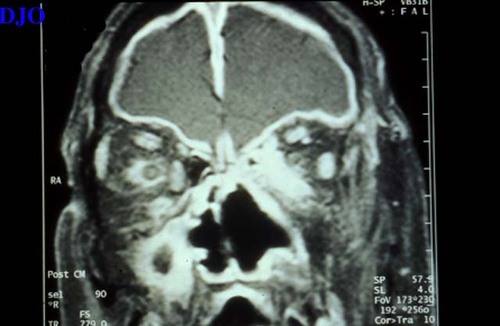
Computed Tomography: see Figures 5-7
Pathology
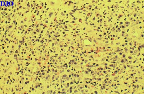
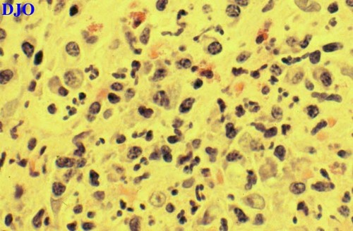
The patient underwent left orbital biopsy: see Figures 8-9 | |
|
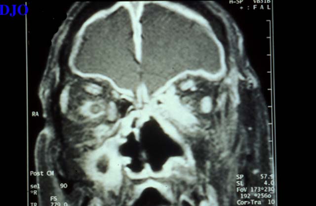
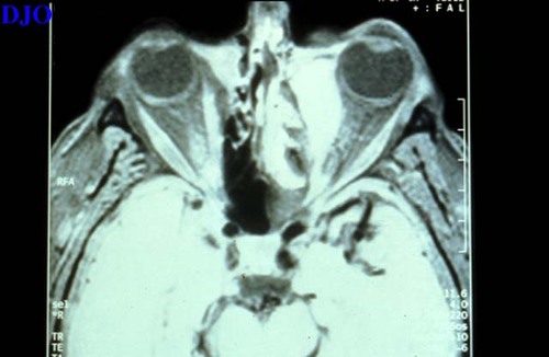
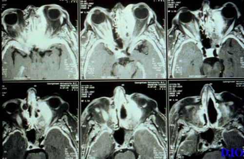
Figure 5
Figures 5-7. Axial and coronal CT scans showing a space occupying infiltrative lesion extending throughout the medial aspect of the left orbit and extending to infiltrate the meninges of the optic nerve bilaterally in the extra-orbital space.
 |
|
|
Figure 6
 |
|
|
Figure 7
 |
|
|
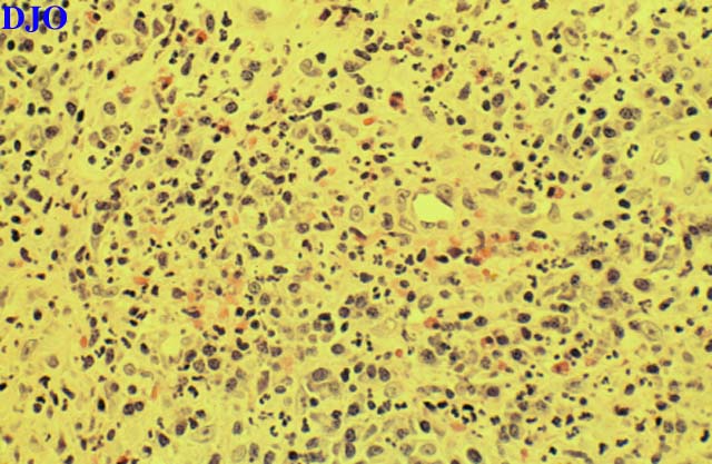
Figure 8
Figures 8-9. Low and high power microscopic views showing necrotizing granulomatous inflammation.
 |
|
|
Figure 9
 |
|
|
|
|
|
 Welcome, please sign in