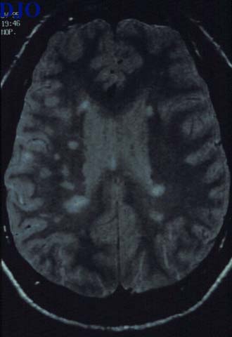
Figure 5
MRI of brain: T2 weighted axial image showing many bright lesions in the periventricular white matter.
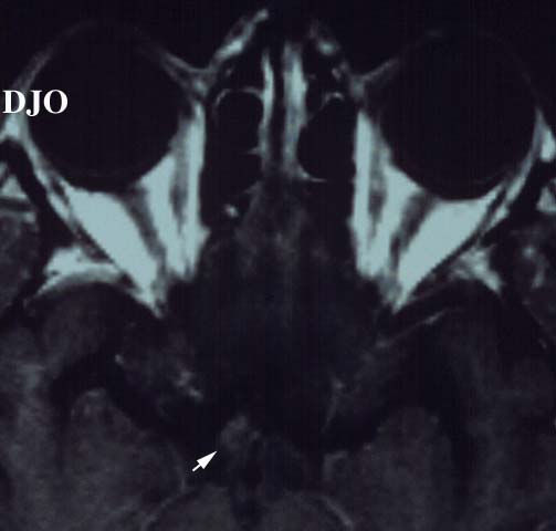
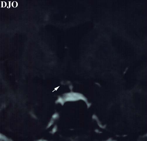
Figure 6
Figures 6-7. Brain MRI: Axial image showing the optic tract: There is enhancement and slight enlargement of the posterior right chiasm and optic tract. Coronal image of the right optic tract also shows enhancement.
 Welcome, please sign in