|
|
 |
|
|
|
|
|
A 64-year-old man with an unusual conjunctival cyst
Digital Journal of Ophthalmology 2017
Volume 23, Number 1
March 8, 2017
DOI: 10.5693/djo.03.2016.04.001
|
Printer Friendly
Download PDF |
|
|

Seanna Grob, MD, MAS | Department of Ophthalmology, Veterans Affairs Boston Healthcare System, Boston, Massachusetts Department of Ophthalmology, Harvard Medical School, Boston, Massachusetts Department of Ophthalmology, Massachusetts Eye and Ear Infirmary Daniel R. Lefebvre, MD | Department of Ophthalmology, Veterans Affairs Boston Healthcare System, Boston, Massachusetts Department of Ophthalmology, Harvard Medical School, Boston, Massachusetts Department of Ophthalmology, Massachusetts Eye and Ear Infirmary Ophthalmic Plastic Surgery, Massachusetts Eye and Ear Infirmary, Boston, Massachusetts Nora Laver, MD | Ophthalmic Pathology, Department of Ophthalmology, Tufts Medical Center and Tufts University School of Medicine, Massachusetts Mary K. Daly, MD | Department of Ophthalmology, Veterans Affairs Boston Healthcare System, Boston, Massachusetts Department of Ophthalmology, Boston University School of Medicine, Boston, Massachusetts Department of Ophthalmology, Harvard Medical School, Boston, Massachusetts
|
|
|
| Ancillary Testing | Cataract surgery was deferred, and the patient underwent excision of the medial canthus conjunctival cyst for diagnostic purposes.
Histopathological Analysis
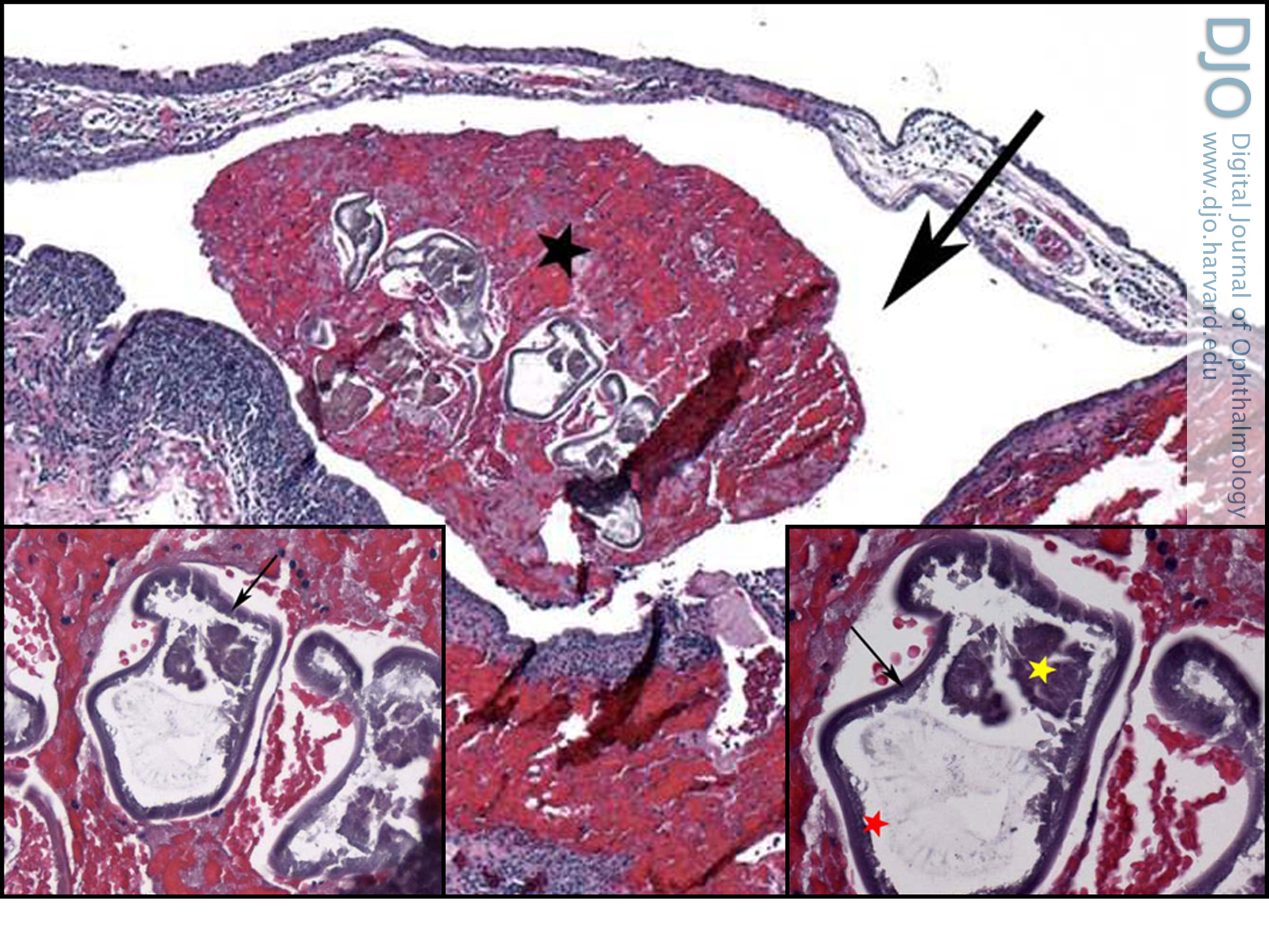
Paraffin-embedded, formalin-fixed tissue slides showed a chronically inflamed conjunctival cyst. (Figure 2). Cross-sections of the cyst revealed a degenerating nematode (Figure 2A-C). Sections through the degenerating nematode showed smooth, refractile cuticles, internal lateral ridges, and a suggestion of degenerated internal organs (Figure 2). Pathology images were sent to the Center of Disease Control (CDC), which confirmed the specimen was a nematode but was unable to prove any further classification. | |
|
Figure 2
Histopathological analysis (hematoxylin and eosin, original magnification ×4): subconjunctival cyst (black arrow), with degenerating nematode particles (black asterisk). Inset left, higher magnification of the degenerating nematode (×20). Inset right, higher magnification (×40) of the degenerating nematode, with smooth, refractile cuticles (black arrow), internal lateral ridges (red asterisk), and a suggestion of degenerated internal organs (yellow asterisk).
 |
|
|
|
|
|
|
|
 Welcome, please sign in