|
|
 |
|
|
|
|
|
49 year old man with 6 months of swelling around the right eye
Digital Journal of Ophthalmology 1998
Volume 4, Number 20
July 6, 1998
|
Printer Friendly
|
|
|
|
|
|
|
| Examination | Vision cc: 20/40 OD, 20/25 OS
External: 2 mm of proptosis OD with Hertels
Pupils: Equal, briskly reactive to light. No RAPD
Extraocular Movements: Normal OU
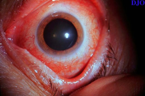
Slit lamp examination OD: 2+ conjunctival injection, cornea clear, Anterior chamber shallow, with trace white blood cells, the iris showed 2 patent peripheral iridectomies.
Slit lamp examination OS: Normal with deep anterior chamber
Gonioscopy: Shallow OD, but open angle. OS angle is wide open
Intraocular pressure: 20 OD, 14 OS
Fundus examination: OD see below, Normal OS | |
|
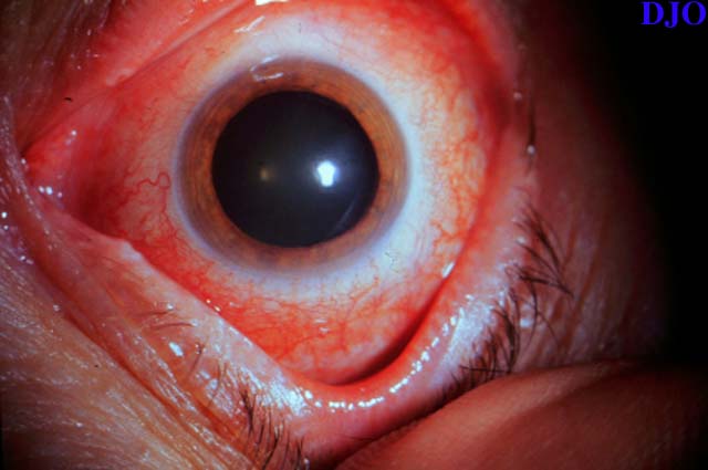
Figure 1
Anterior segment examination showing a diffusely injected conjunctiva. The cornea was clear, the anterior chamber shallow with 1+ white blood cells.
 |
|
|
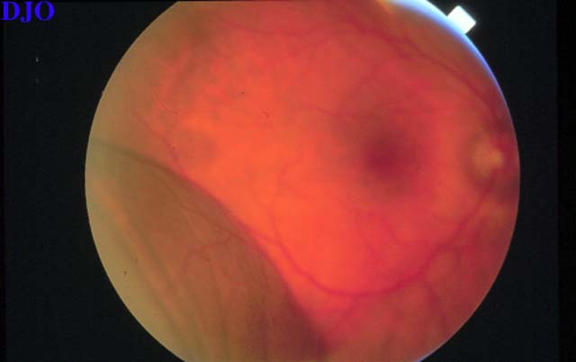
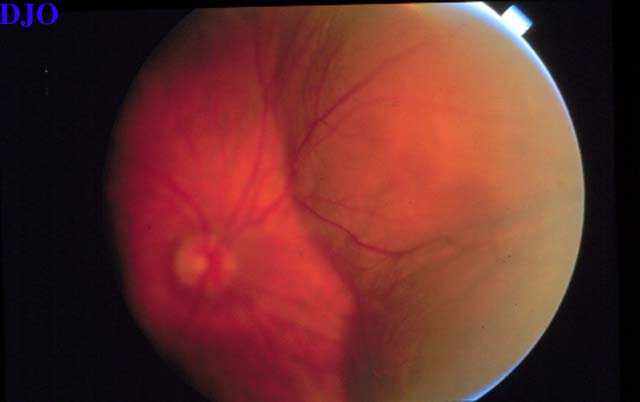
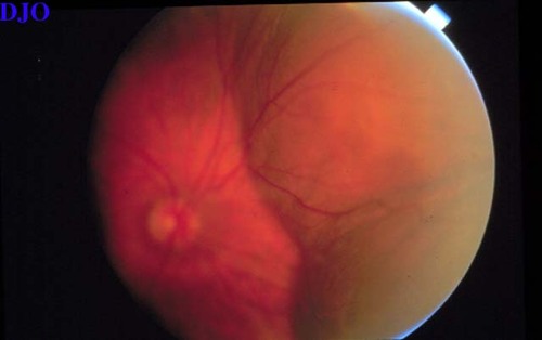
Figure 2
Figures 2-3. Fundoscopic examination of the right eye revealing an elevated mass in the inferotemporal quadrant and the nasal half of the retina. There was no retinal detachment or vitritis. The fluid did not shift with position change. Examination of the far periphery showed 360 degree cilio-choroidal detachment in an annular configuration.
 |
|
|
Figure 3
 |
|
|
|
|
|
|
|
 Welcome, please sign in