|
|
 |
|
|
|
|
|
A 24-year-old woman with rapidly progressing vision loss
Digital Journal of Ophthalmology 2017
Volume 23, Number 1
January 15, 2017
DOI: 10.5693/djo.03.2016.10.001
|
Printer Friendly
Download PDF |
|
|
 Milad Modabber, MD, MSc
 | Department of Ophthalmology, McGill University, Montreal, Quebec, Canada Vasudha Gupta MD, FRCSC | Department of Ophthalmology, Queen’s University, Kingston, Ontario, Canada Amadeo R. Rodriguez, MD | Department of Surgery Ophthalmology and Medicine Neurology, McMaster University, Hamilton, Ontario, Canada
|
|
|
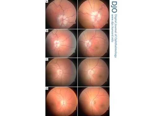
| Examination | | On initial examination, the patient’s best-corrected visual acuity was 20/100 in the right eye and 20/70 in the left eye. The pupils were symmetrically 3 mm in diameter and poorly responsive to light in both eyes, with no evidence of a relative afferent pupillary defect. Intraocular pressure was recorded as 16 mm Hg in each eye. Slit-lamp biomicroscopy was unremarkable. Dilated fundus examination revealed marked optic nerve head swelling suggestive of papilledema and tortuous and dilated retinal venules (Figure 1A). | |
|
Figure 1
Fundus images of the right eye (left column) and left eye (right column) at acute presentation (A), and at 1 week (B), 6 weeks (C), and 18 months (D) after surgery.
 |
|
|
|
|
|
|
|
 Welcome, please sign in


