|
|
 |
|
|
|
|
|
A 24-year-old woman with rapidly progressing vision loss
Digital Journal of Ophthalmology 2017
Volume 23, Number 1
January 15, 2017
DOI: 10.5693/djo.03.2016.10.001
|
Printer Friendly
Download PDF |
|
|
 Milad Modabber, MD, MSc
 | Department of Ophthalmology, McGill University, Montreal, Quebec, Canada Vasudha Gupta MD, FRCSC | Department of Ophthalmology, Queen’s University, Kingston, Ontario, Canada Amadeo R. Rodriguez, MD | Department of Surgery Ophthalmology and Medicine Neurology, McMaster University, Hamilton, Ontario, Canada
|
|
|
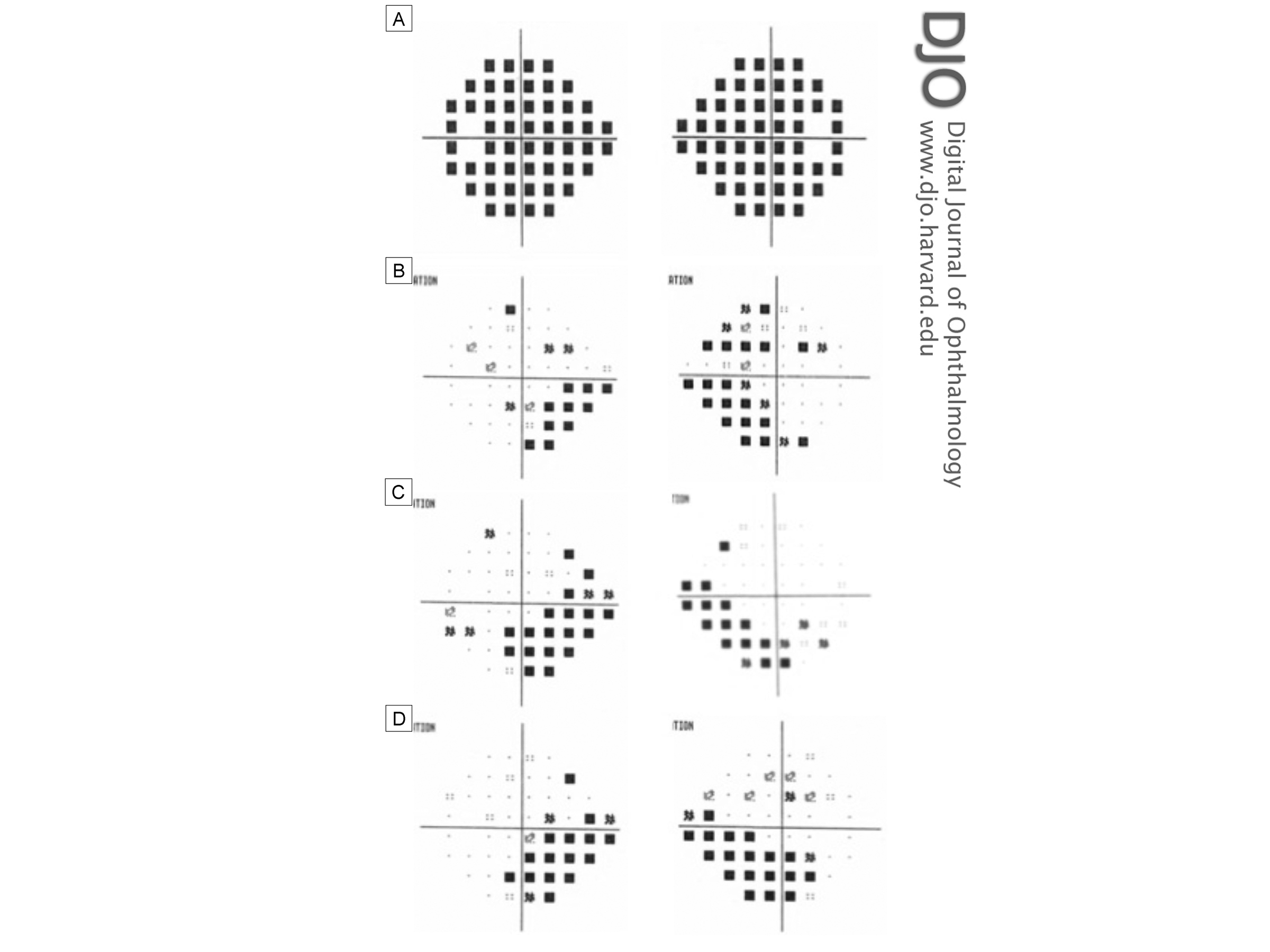
| Ancillary Testing | | An initial head computed tomography (CT) scan was unremarkable. Lumbar puncture demonstrated an elevated opening pressure of 60 cm H2O. The cerebrospinal fluid (CSF) composition was normal. Magnetic resonance imaging (MRI) of the head revealed increased perioptic space around both optic nerves with flattening of the posterior sclera suggestive of increased intracranial pressure but was otherwise unremarkable. Magnetic resonance venography (MRV) was normal, with no evidence of dural sinus thrombosis. Visual field assessment (Humphrey 24-2 demonstrated severe and diffuse visual field loss bilaterally with preservation of central vision, with a mean deviation of −32.0 dB in the right eye and −28.6 dB in the left eye (Figure 2A; pattern deviation not shown due to severely depressed fields). | |
|
Figure 2
Humphrey’s 24-2 visual field total and/or pattern deviation results of the right eye (right column) and left eye (left column). A, Total deviation at acute presentation (pattern deviations not shown due to severely depressed fields). B, Pattern deviation at 6 weeks after surgery. C, Pattern deviation at 4 months after surgery. D, Pattern deviation at 1 year after surgery
 |
|
|
|
|
|
|
|
 Welcome, please sign in