|
|
 |
|
|
|
|
|
45 year old lady who complained of decreased vision OS of one year duration after cataract surgery
Digital Journal of Ophthalmology 1998
Volume 4, Number 19
June 9, 1998
|
Printer Friendly
|
|
|

DV Inglesby MB, BSc, FRCS, FRCOphth | Sunderland Eye Infirmary, Sunderland, United Kingdom LM Tong MB, BS | Sunderland Eye Infirmary, Sunderland, United Kingdom
|
|
|
| Examination | Vision: 6/9 OD, 6/36 OS
Pupils: Equal, reactive, No APD
Motility: Full OU
External: No lid swelling or conjunctival chemosis or injection.
Intraocular pressure: 16 OD, 24 OS
Slit lamp examination: see Figure 1
Dilated fundus examination: Normal OD | |
|
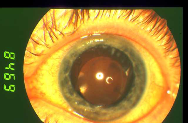
Figure 1
The left eye showed a few cells in the anterior chamber. The anterior segment otherwise showed no obvious inflammation. The cornea and previous incision site looked normal. The pupil was round. A posterior chamber intraocular lens implant was visualised in the capsular bag. There was posterior capsular opacification with a small opening centrally. Peripheral fluffy white spots could be seen superiorly.
 |
|
|
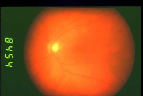
Figure 2
Here is the fundus photo of the patient's left eye. The view was hazy. A few pigmented cells could be seen in the anterior vitreous. The macula looked thickened with binocular fundoscopy, but this is not apparent on the colour photograph. The optic disc was normal.
 |
|
|
|
|
|
|
|
 Welcome, please sign in