|
|
 |
|
|
|
|
|
52 year old Filipino female with decreased vision OD for two weeks
Digital Journal of Ophthalmology 1998
Volume 4, Number 17
April 18, 1998
|
Printer Friendly
|
|
|

Everett Ai, MD | California Pacific Medical Center, San Francisco, CA Haris I. Amin, MD | California Pacific Medical Center, San Francisco, CA
|
|
|
| Ancillary Testing | Angiogram
| |
|
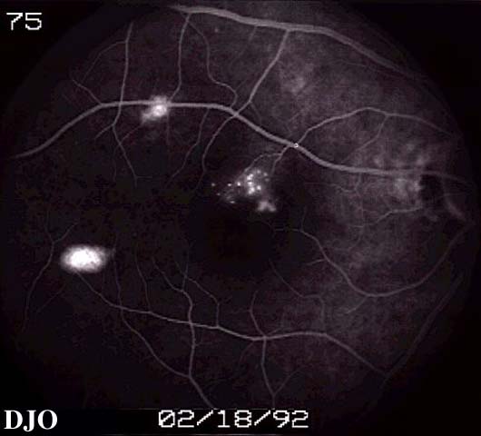
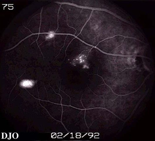
Figure 4
Angiogram of the right eye FROM 1992 shows window defects. Laser photocoagulation had been performed twice before.
 |
|
|
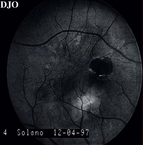
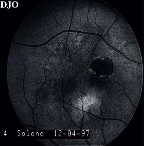
Figure 5
Red free photo of the right eye shows scar and subretinal fluid. Also note the gray streak-like lesions in the choroid peripherally.
 |
|
|
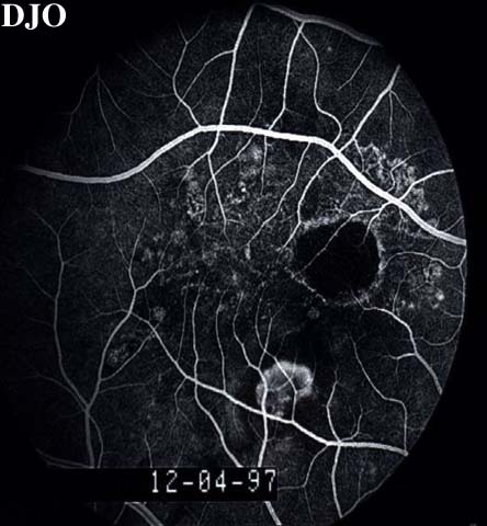
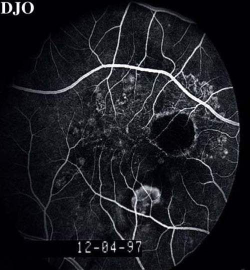
Figure 6
Laminar venous phase angiogram of OD shows blocked fluorescence FROM the scar, hyperfluorescent lesion inferior to the fovea, and peripheral hyperfluorescence corresponding to the streak-like gray choroidal lesions.
 |
|
|
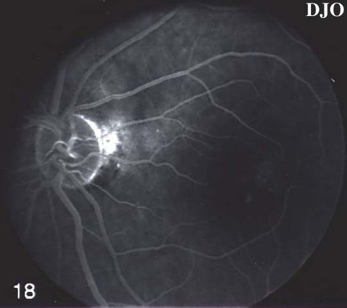
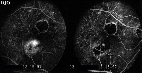
Figure 7
Angiogram of OS shows hyperfluorescence in the macula.
 |
|
|
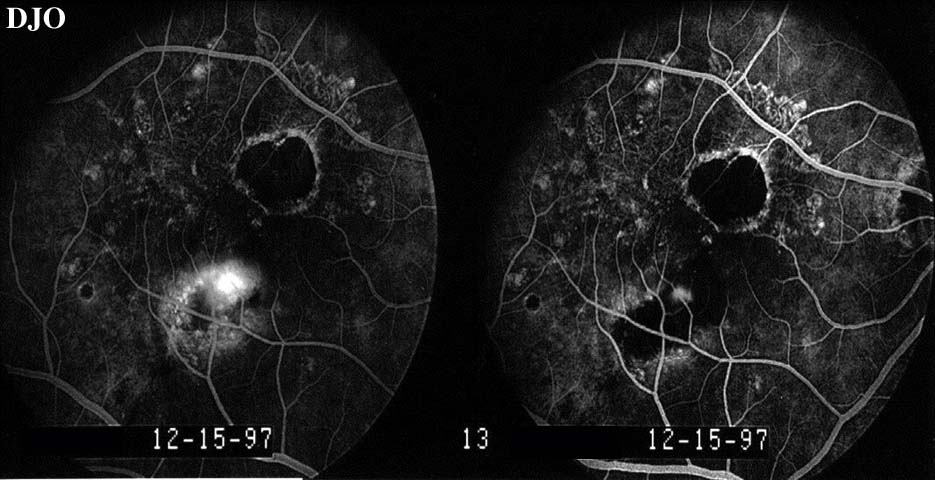
Figure 8
The lesion in the right eye was treated with laser photocoagulation. Acuity improved to 20/40 FROM 20/50. This figure shows the followup angiogram. There is still some leakage INTO the lesion which was retreated.
 |
|
|
|
|
|
|
|
 Welcome, please sign in