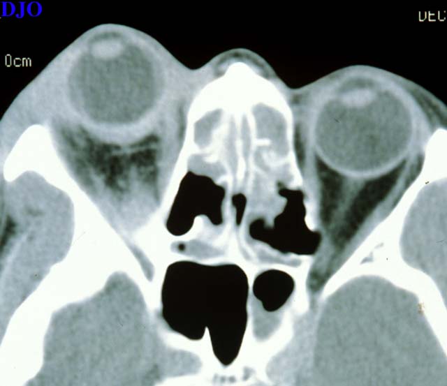
Figure 3a
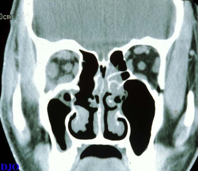
Figures 3a-3b. Orbital CT (Left) Axial, showing periorbital soft tissue swelling, thickening of the posterior sclera, choroidal elevations, and proptosis OD. (Right) Coronal, showing thickening of the extraocular muscles OD.
Figure 3b
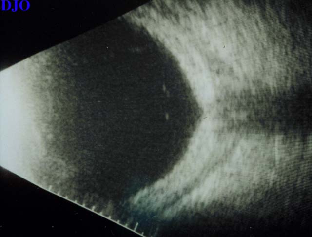
Figure 4a
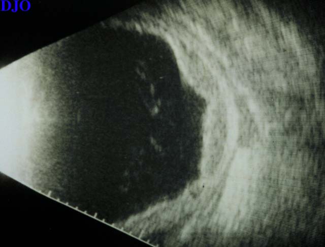
Figures 4a-4b. B-scan ultrasound (Left) Posterior, showing a sub-tenon's effusion at the junction of the optic nerve and globe (T-sign). (Right) Temporal, showing an exudative choroidal effusion and a sub-tenon's effusion.
 Welcome, please sign in