|
|
 |
|
|
|
|
|
77 year old woman seen in the optometry clinic for aphakic contact lenses
Digital Journal of Ophthalmology 1998
Volume 4, Number 15
February 28, 1998
|
Printer Friendly
|
|
|

Kenneth M. Goins, MD | The University of Chicago, Chicago IL Arutun Oganesian, MD | The University of Chicago, Chicago IL Louise Sclafani, OD | The University of Chicago, Chicago IL
|
|
|
| Examination | Vision: 20/40 OD; 20/30 OS with contact lenses OU
External exam: Normal
Pupils: Reactive to light, no APD OU
Motility: Full OU
Slit lamp examination: See figures 1-3
Endothelial Specular Microscopy: See figures 4-6
Intraocular pressure: 14 mm Hg OD, 14 mm Hg OS
Fundi: Normal OU | |
|
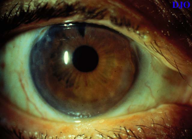
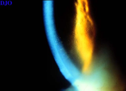
Figure 1
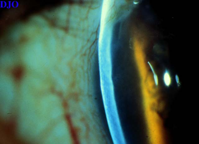
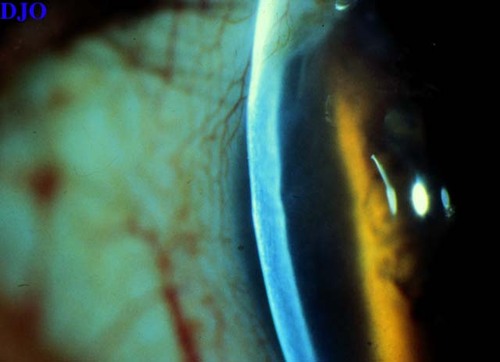
Figures 1-3. There is peripheral concentric corneal edema extending 2.5-3.5 mm FROM the limbus. The patient has a peripheral iridectomy and is aphakic OD. The left eye had a similar appearance.
 |
|
|
Figure 2
 |
|
|
Figure 3
 |
|
|
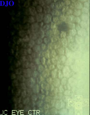
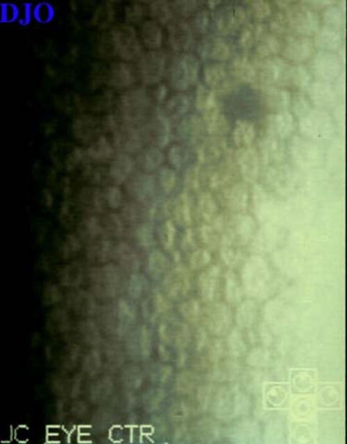
Figure 4
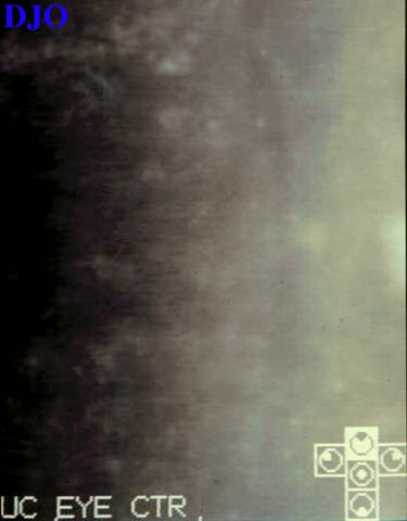
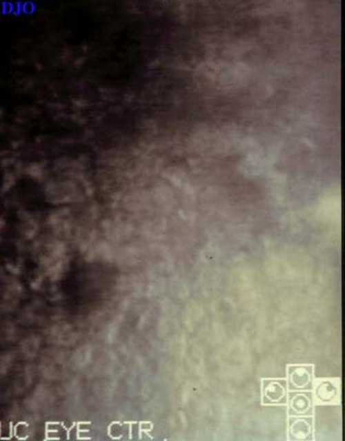
Figures 4-6. Specular microscopy of the center of the cornea is essentially normal(left), Specular microscopy of the peripheral cornea (center and right) shows polymegathism and decreased numbers of corneal endothelial cells
 |
|
|
Figure 2
 |
|
|
Figure 6
 |
|
|
|
|
|
|
|
 Welcome, please sign in