Sarper Karaküçük, MD | Erciyes University Faculty of Medicine, Kayseri, Turkey G. Ertugrul Mirza, M.D. | Erciyes University Faculty of Medicine, Kayseri, Turkey
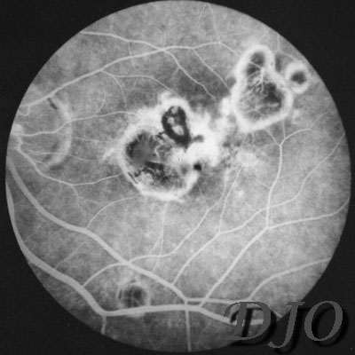
Figure 3a
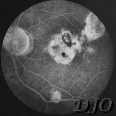
Figures 3a-3b, Florescein angiogram (OS). Fluorescein angiogram showing areas of hyperfluorescence (a) which increases at the late phase(b); window defects as well as atrophic scars on the left macular region are seen.
Figure 3b
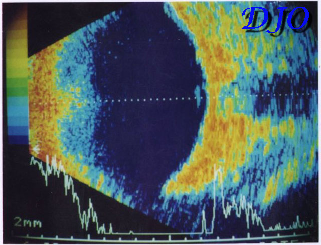
Figure 4
Ultrasonography (OS). PVD is seen as a vertical line at the posterior fundus.
 Welcome, please sign in