|
|
 |
|
|
|
|
|
A 45-year-old man with spontaneous hyphema of the right eye
Digital Journal of Ophthalmology 2017
Volume 23, Number 3
August 9, 2017
DOI: 10.5693/djo.03.2017.02.003
|
Printer Friendly
Download PDF |
|
|
 Jay C. Wang, MD
 | Department of Ophthalmology, Massachusetts Eye and Ear, Boston, Massachusetts Maggie B. Hymowitz, MD | Department of Ophthalmology, Massachusetts Eye and Ear, Boston, Massachusetts
|
|
|
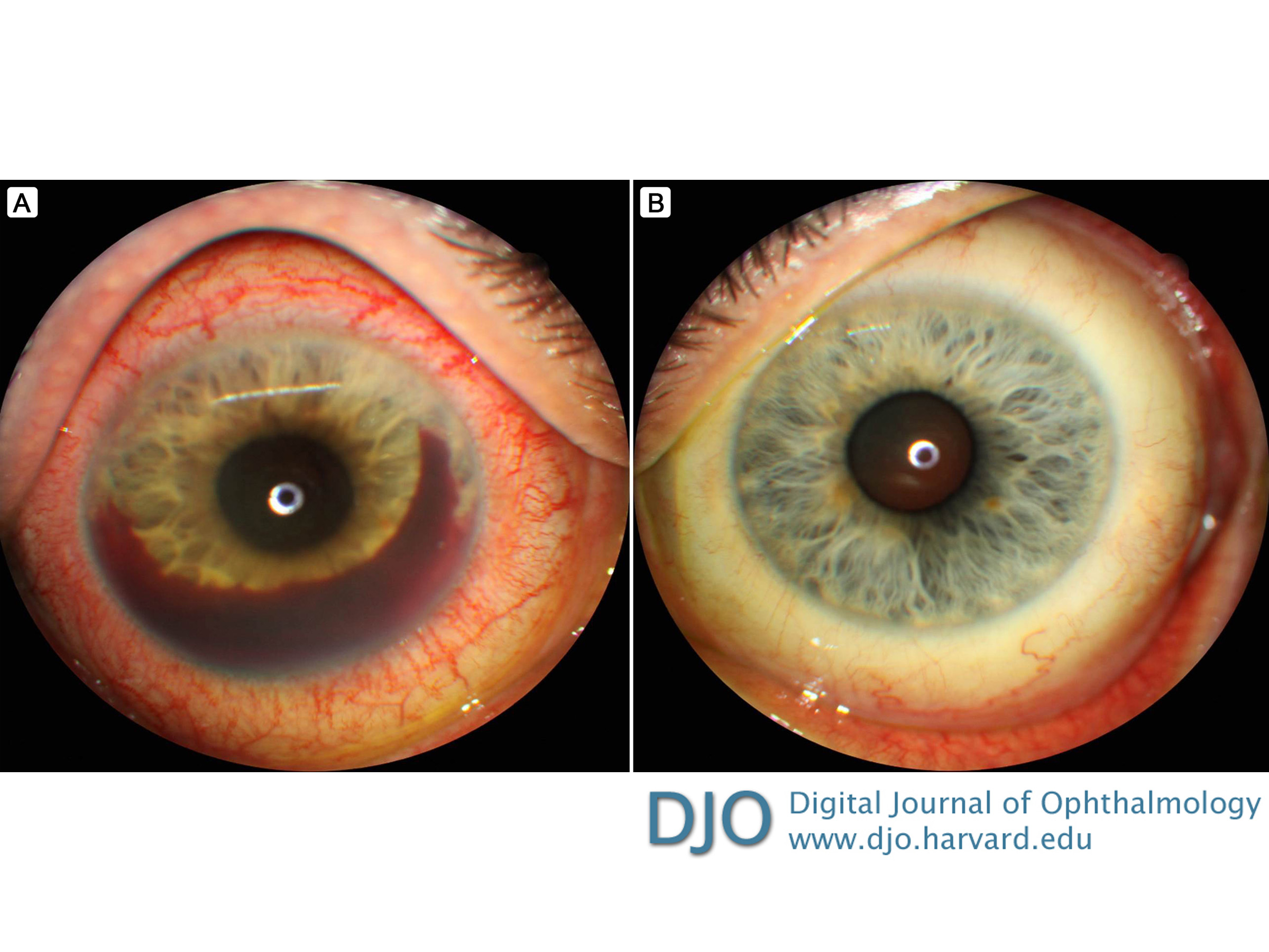
| Examination | On examination, visual acuity was 20/50 in the right eye and 20/20 in the left eye. His pupils were equal, round, and reactive; there was no relative afferent pupillary defect. Intraocular pressure was 18 mm Hg in the right eye and 16 mm Hg in the left eye. Anterior segment examination of the right eye revealed 2+ diffuse conjunctival and scleral injection, with tenderness, fine keratic precipitates, diffuse anterior stromal haze of the cornea, and Descemet folds centrally. Prominent iris blood vessels were noted superiorly with 360 degrees of posterior synechiae, but there were no iris transillumination defects. The anterior chamber was notable for 4+ cell and flare and a 1 mm inferior clot, with 2 mm layering hyphema from 2 to 9 o’clock (Figure 1A). Gonioscopy showed fine blood vessels traversing the superior angle, which did not cross over the scleral spur onto the trabecular meshwork. There were no peripheral anterior synechiae. Posterior segment examination of the right eye was somewhat limited because of the anterior segment pathology and poor pupillary dilation. Only a small, intraretinal hemorrhage inferior to the optic nerve was noted. There was no evidence of vitritis or pars planitits. Anterior segment examination (Figure 1B) and gonioscopy of the left eye were normal. Posterior segment examination of the left eye was unremarkable.
| |
|
Figure 1
Anterior segment photographs of the right eye (A) and the left eye (B).
 |
|
|
|
|
|
|
|
 Welcome, please sign in