|
|
 |
|
|
|
|
|
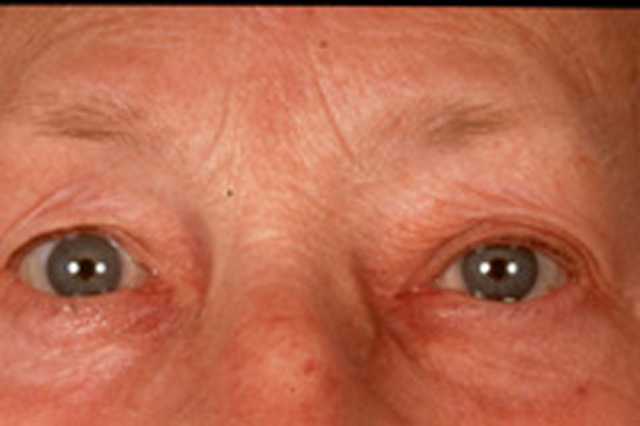
79 year old female with redness and swelling of the left eyelids
Digital Journal of Ophthalmology 2002
Volume 8, Number 3
March 1, 2002
|
Printer Friendly
|
|
|

Aljoscha S Neubauer, M.D. | Ludwig-Maximilians-Universität, 80336 Munich, Germany Christos Haritoglou, M.D. | Ludwig-Maximilians-Universität, 80336 Munich, Germany Christoph Hintschich, M.D. | Ludwig-Maximilians-Universität, 80336 Munich, Germany
|
|
|
| Diagnosis and Discussion | Orbital Abscess
Clinical Course:
The patient was operated on the same day by ENT service as the orbital abscess originated FROM the frontal sinus. The abscess was drained, the i.v. antibiotics were continued.
Four days later the patient was seen as follow-up. Motility was full OU, only slight pain and slight swelling were present. On Hertel no proptosis was measured.
Discussion:
Orbital cellulitis results secondarily FROM acute or chronic bacterial sinusitis in more than 90% of cases. CT scanning to evaluate the paranasal sinuses therefore is essential. Causes of orbital cellulitis include extension FROM periorbital structures (paranasal sinuses, face and eyelids, dacryocystitis, dental infection, intracranial), exogenous causes (trauma, postsurgical), endogenous septic embolization and intraorbital causes (endophthalmitis, dacryoadenitis) (1).
Antibiotics should provide coverage to multiple organisms such as gram-positive cocci, H influenzae, and anaerobes. Abscess formation is not infrequently seen and should usually be drained surgically.
In the presented case two exceptional features were encountered:
1) a very benign clinical presentation of a relatively large orbital abscess
2) difficulty of ultrasonic diagnosis of the abscess
A wide spectrum of clinical presentation of orbital abscesses exist (2), depending on localization, species of bacteria, and immune response of the patient. Age of the patient seems to be an important factor (3), with subperiostal abscesses of younger children more often clearing without drainage. Therapeutic regimens therefore have to depend on the clinical situation (4). Surgical drainage is not always necessary.
Ultrasound and CT are the two main diagnostic studies for imaging an orbita abscess. Orbital complications of inflammatory paranasal sinus diseases can be correctly diagnosed by ultrasound alone in 80 to 100% of the cases (5). Several studies indicate the high specifity and sensitivity of ultrasound diagnosis, especially when differentiating between orbital cellulitis and subperiosteal abscess (6) or when differentiating between cellulitis and pseudotumor (7). However, for localization of orbital abscesses the CT still remains the vital imaging study (7), especially when localizing the sinusitis causing the abscess. Therefore both imaging studies - CT and ultrasound - should be applied to patients, when an orbital abscess is suspected. | |
|
Figure 5
 |
|
|
|
|
|
|
|
 Welcome, please sign in