|
|
 |
|
|
|
|
|
72 year old male with progressive bilateral decrease in vision
Digital Journal of Ophthalmology 2002
Volume 8, Number 4
May 1, 2002
|
Printer Friendly
|
|
|

Manoj M. Thakker, MD | Massachusetts Eye & Ear Infirmary, Boston MA Deborah S. Jacobs, MD | Beth Israel/Deaconess Medical Center, Boston MA
|
|
|
| Ancillary Testing | Pathology
The patient underwent a transsphenoidal resection of the pituitary mass. | |
|
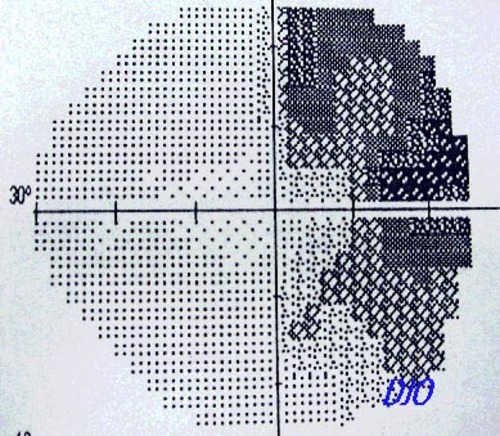
Figure 1a
Figures 1a-1b. Humphrey visual field reveals a bitemporal hemianopia
 |
|
|
Figure 1b
 |
|
|
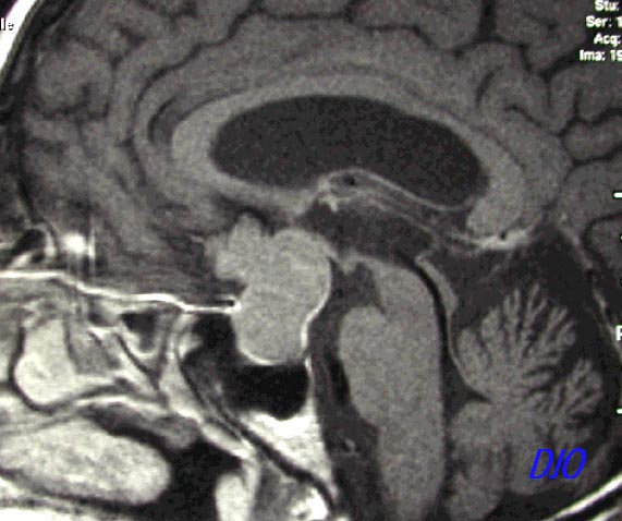
Figure 2a
Figures 2a - 2b. MRI shows a homogeneous mass extending superiorly FROM the pituitary fossa INTO the third ventricle and anterior cranial fossa.The mass extends superiorly FROM the pituitary fossa INTO the third ventricle and laterally toward the cavernous sinuses
 |
|
|
Figure 2b
 |
|
|
Figure 3a
Figures 3a-3b. Pathology revealed a pituitary adenoma which did not immunoreact with antibodies to ACTH, FSH, LH, TSH, prolactin, S-100 (marker for melanoma and neuroectodermal tumors), or L-26 (B-cell marker)
 |
|
|
Figure 3b
 |
|
|
|
|
|
 Welcome, please sign in