|
|
 |
|
|
|
|
|
A 73-year-old man with congestion and mild proptosis of the left eye
Digital Journal of Ophthalmology 2016
Volume 22, Number 2
April 25, 2016
DOI: 10.5693/djo.03.2014.05.001
|
Printer Friendly
Download PDF |
|
|
 Amanda Mohanan Earatt, MS
 | Department of Ophthalmology, Amala Institute of Medical Sciences, Thrissur, Kerala, India Lathika Vasu Kamaladevi, MS, DO, FRCS Glasgow | Department of Ophthalmology, Amala Institute of Medical Sciences, Thrissur, Kerala, India Charles K. Skariah, MS, DO | Department of Ophthalmology, Amala Institute of Medical Sciences, Thrissur, Kerala, India
|
|
|
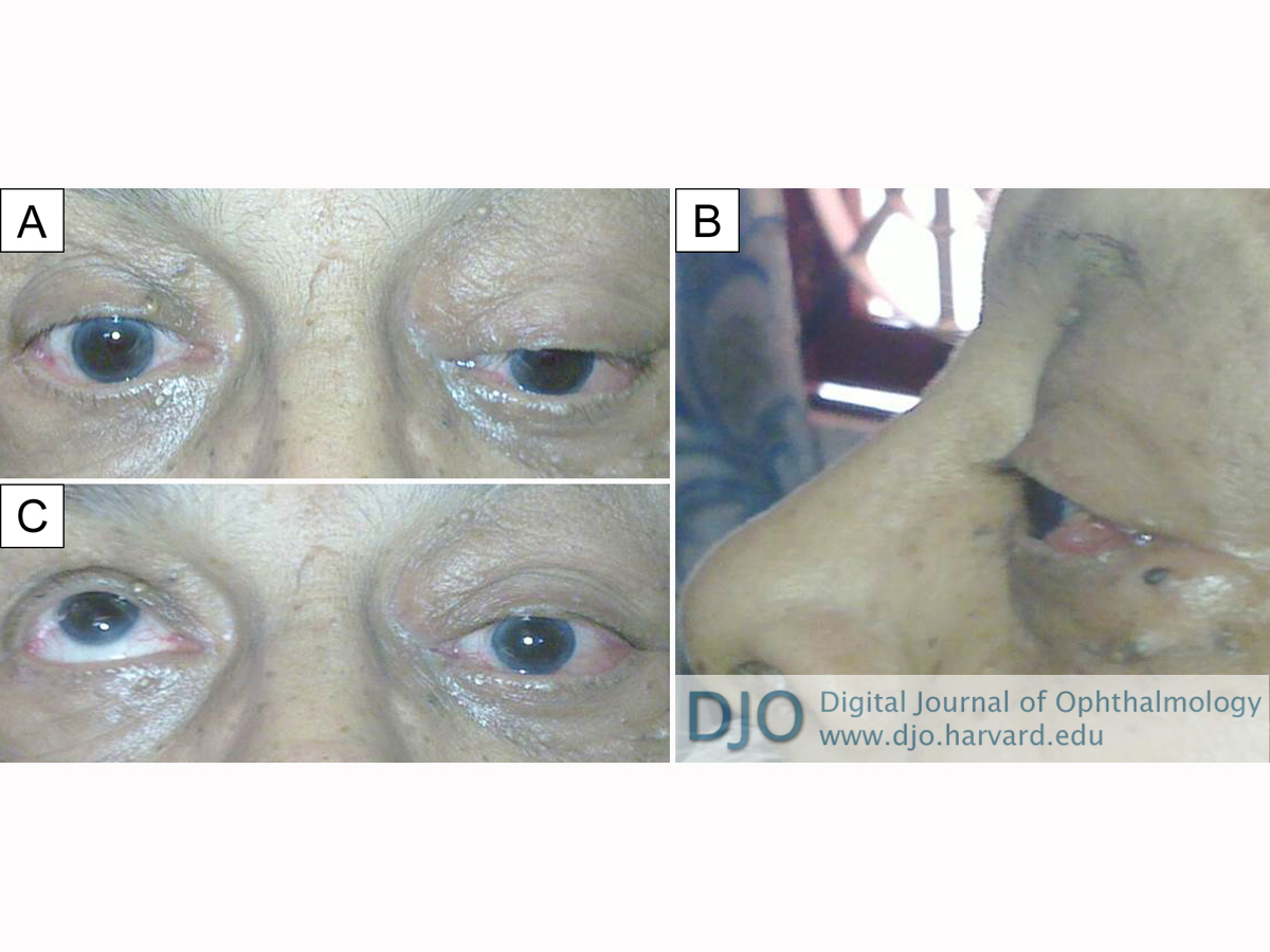
| Examination | On initial examination, best-corrected visual acuity was 20/20 in each eye. Color vision was normal in both eyes. There was periorbital fullness and partial ptosis of the left eye. Palpation revealed a painless, firm swelling along the lateral one-third of superior orbital margin. It was found to displace the globe inferiorly (Figure 1A). Hertel exophthalmometry measured 16 mm in the right eye and 22 mm in the left eye (Figure 1B). Ocular motility was full on the right side; the left eye showed generalized restriction and complete loss of elevation (Figure 1C). Slit-lamp examination of the anterior and posterior segments was entirely normal except for temporal bulbar conjunctival congestion with chemosis in left eye. Pupils were reactive, with no afferent pupillary defect.
General examination did not reveal any abnormal cervical lymph nodes. Cranial nerve evaluation was within normal limits. | |
|
Figure 1
Clinical photograph of the patient at presentation. A, Left periorbital fullness and partial ptosis, with inferior displacement of the globe. B, Proptosis of left eye with temporal conjunctival congestion and chemosis. C, Complete loss of elevation in left eye.
 |
|
|
|
|
|
|
|
 Welcome, please sign in