|
|
 |
|
|
|
|
|
A 73-year-old man with congestion and mild proptosis of the left eye
Digital Journal of Ophthalmology 2016
Volume 22, Number 2
April 25, 2016
DOI: 10.5693/djo.03.2014.05.001
|
Printer Friendly
Download PDF |
|
|
 Amanda Mohanan Earatt, MS
 | Department of Ophthalmology, Amala Institute of Medical Sciences, Thrissur, Kerala, India Lathika Vasu Kamaladevi, MS, DO, FRCS Glasgow | Department of Ophthalmology, Amala Institute of Medical Sciences, Thrissur, Kerala, India Charles K. Skariah, MS, DO | Department of Ophthalmology, Amala Institute of Medical Sciences, Thrissur, Kerala, India
|
|
|
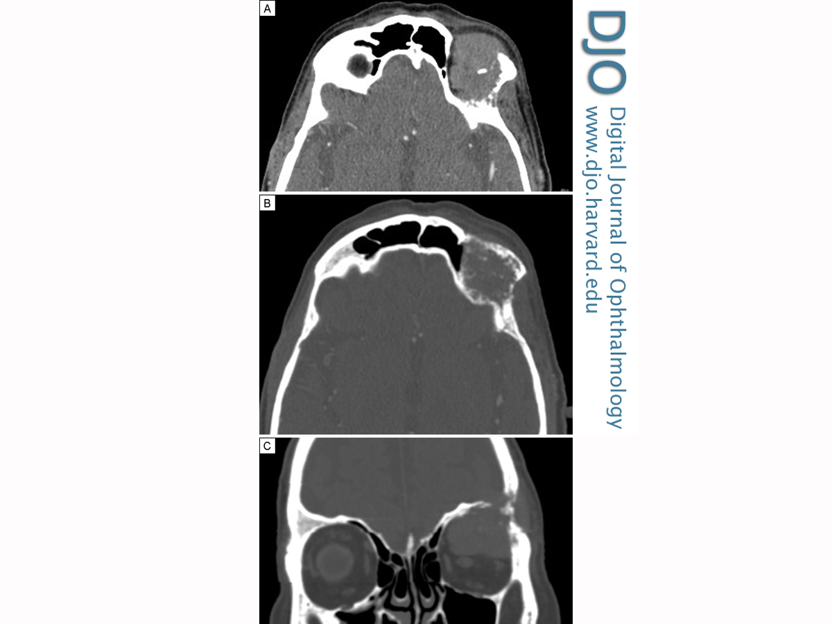
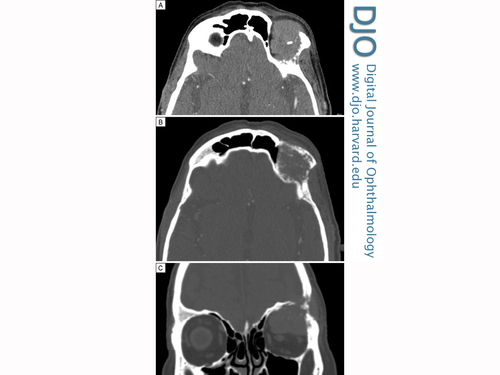
| Ancillary Testing | Routine blood testing, thyroid function tests, and prostate specific antigen (PSA; 0.1 ng/ml) were within normal limits. Computed tomography (CT) of brain and orbit demonstrated an enhancing lytic lesion, with a significant soft tissue component and calcification. It measured 22 × 35 × 34 mm and involved the left frontal bone encroaching on the lateral wall of orbit. The lesion was found to indent the globe on its superior aspect. Both the superior rectus and superior oblique muscles appeared to be involved. The left optic nerve was free. These findings were suggestive of left orbital metastasis with intracranial extension (Figure 2).
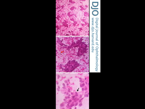
Fine-needle aspiration cytology of the mass was undertaken and a diagnosis of poorly differentiated carcinoma, possibly adenocarcinoma was made (Figure 3). PSA staining confirmed prostatic origin. | |
|
Figure 2
Computed tomography of the brain and orbits. Axial images (A, soft tissue window; B, bone window) showing enhancing lytic lesion (22 × 35 × 34 mm), with significant soft tissue component and calcification. Coronal reconstruction (C) showing the lesion eroding left frontal bone on the lateral wall of the orbit.
 |
|
|
Figure 3
Fine-needle aspiration cytology showing round to polygonal cells having hyperchromatic pleomorphic nuclei, with indistinct nucleoli and moderate amount of eosinophilic cytoplasm (black arrow); scattered bare nuclei (red arrow) were also noted (H & E, original magnification ×40, ×100, ×400).
 |
|
|
|
|
|
|
|
 Welcome, please sign in