|
|
 |
|
|
|
|
|
A 27-year-old man with traumatic partial dislocation of an intraocular lens
Digital Journal of Ophthalmology 2016
Volume 22, Number 1
February 2, 2016
DOI: 10.5693/djo.03.2015.10.003
|
Printer Friendly
Download PDF |
|
|

Cory Miller, BS | University of Minnesota Medical School, Minneapolis Luke Dolezal, BA | University of Minnesota Medical School, Minneapolis Sandra R. Montezuma, MD | Department of Ophthalmology & Visual Neurosciences. University of Minnesota, Minneapolis
|
|
|
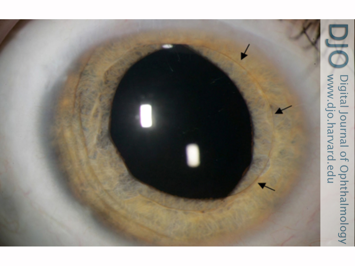
| Examination | | On examination, uncorrected visual acuity in each eye was 20/25. Intraocular pressure (IOP) measured by Tono-Pen (Reichert Inc, Buffalo, NY) was 17 mm Hg in each eye. There was no relative afferent pupillary defect in either eye. Pupillary discoria of the right eye was observed. On slit-lamp examination of the right eye, the conjunctiva, sclera, and cornea were normal. There were a few pigmented cells in the anterior chamber. The optic of the IOL had a partial pupillary capture in which half of the optic was anterior to the iris. The haptics of the lens remained in the sulcus (Figure 1). Anterior segment examination of the left eye was unremarkable and revealed a well-positioned posterior chamber IOL. Fundus examination of both eyes revealed pink optic nerves, flat maculae, and attached retinae. | |
|
Figure 1
Partial pupillary capture of the intraocular lens (IOL). Arrows indicate the edges of the anteriorly dislocated optic.
 |
|
|
|
|
|
|
|
 Welcome, please sign in