|
|
 |
|
|
|
|
|
A 61-year-old man with cystoid macular edema and chorioretinal folds after cataract surgery
Digital Journal of Ophthalmology 2017
Volume 23, Number 3
August 29, 2017
DOI: 10.5693/djo.03.2017.02.002
|
Printer Friendly
Download PDF |
|
|

Andrew Lee, MD | Department of Ophthalmology and Visual Neuroscience, University of Minnesota Sandra R. Montezuma, MD | Department of Ophthalmology and Visual Neuroscience, University of Minnesota
|
|
|
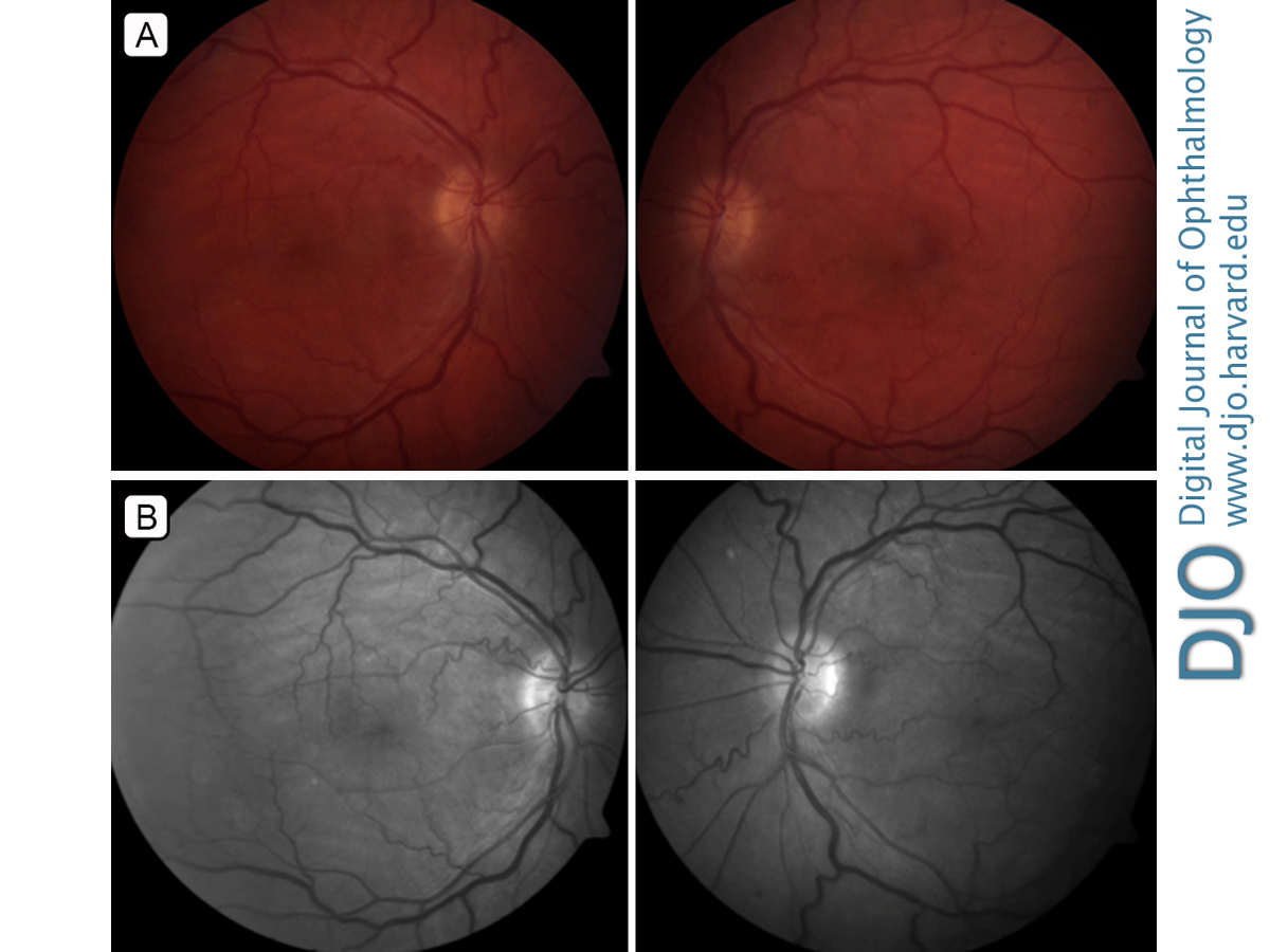
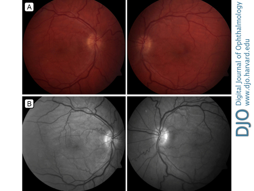
| Examination | | On examination, uncorrected visual acuity was 20/60 in the right eye and 20/40 in the left eye with no pinhole improvement. Intraocular pressure was 19 mm Hg in the right eye and 18 mm Hg in the left eye. Motility was full, with no ptosis or proptosis. Anterior segment examination was unremarkable. A posterior chamber IOL was present and in good position in both eyes. Fundus examination (Figure 1) in each eye showed macular edema and a pink optic nerve, with slightly blurred disk margins. There was mild tortuosity of the vessels, dilation of the venules, and alternating dark and light yellowish streaks in the posterior pole corresponding to chorioretinal folds. | |
|
Figure 1
Fundus photographs (A) and red-free photographs (B) showing chorioretinal folds, venular dilation, and vessel tortuosity.
 |
|
|
|
|
|
|
|
 Welcome, please sign in