|
|
 |
|
|
|
|
|
A 61-year-old man with cystoid macular edema and chorioretinal folds after cataract surgery
Digital Journal of Ophthalmology 2017
Volume 23, Number 3
August 29, 2017
DOI: 10.5693/djo.03.2017.02.002
|
Printer Friendly
Download PDF |
|
|

Andrew Lee, MD | Department of Ophthalmology and Visual Neuroscience, University of Minnesota Sandra R. Montezuma, MD | Department of Ophthalmology and Visual Neuroscience, University of Minnesota
|
|
|
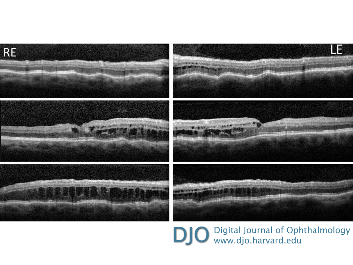
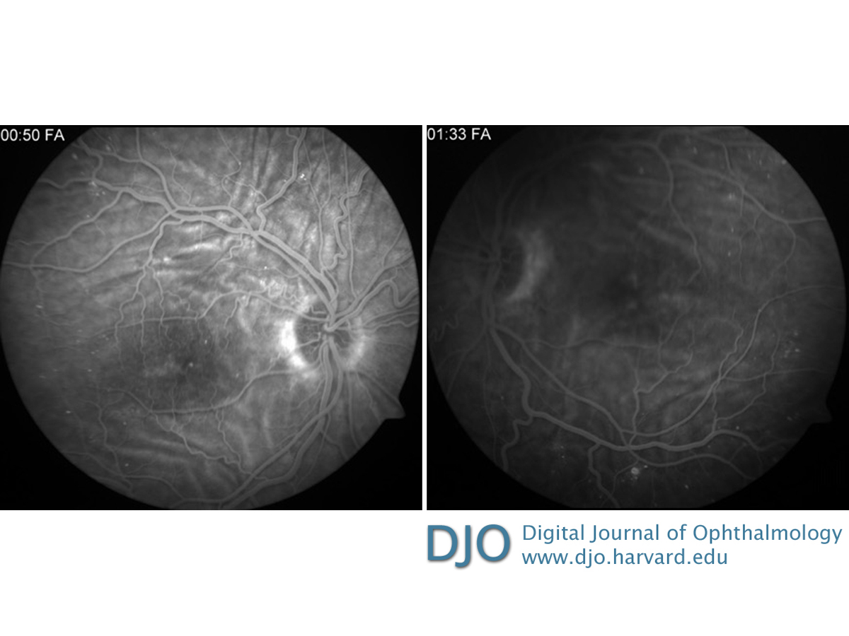
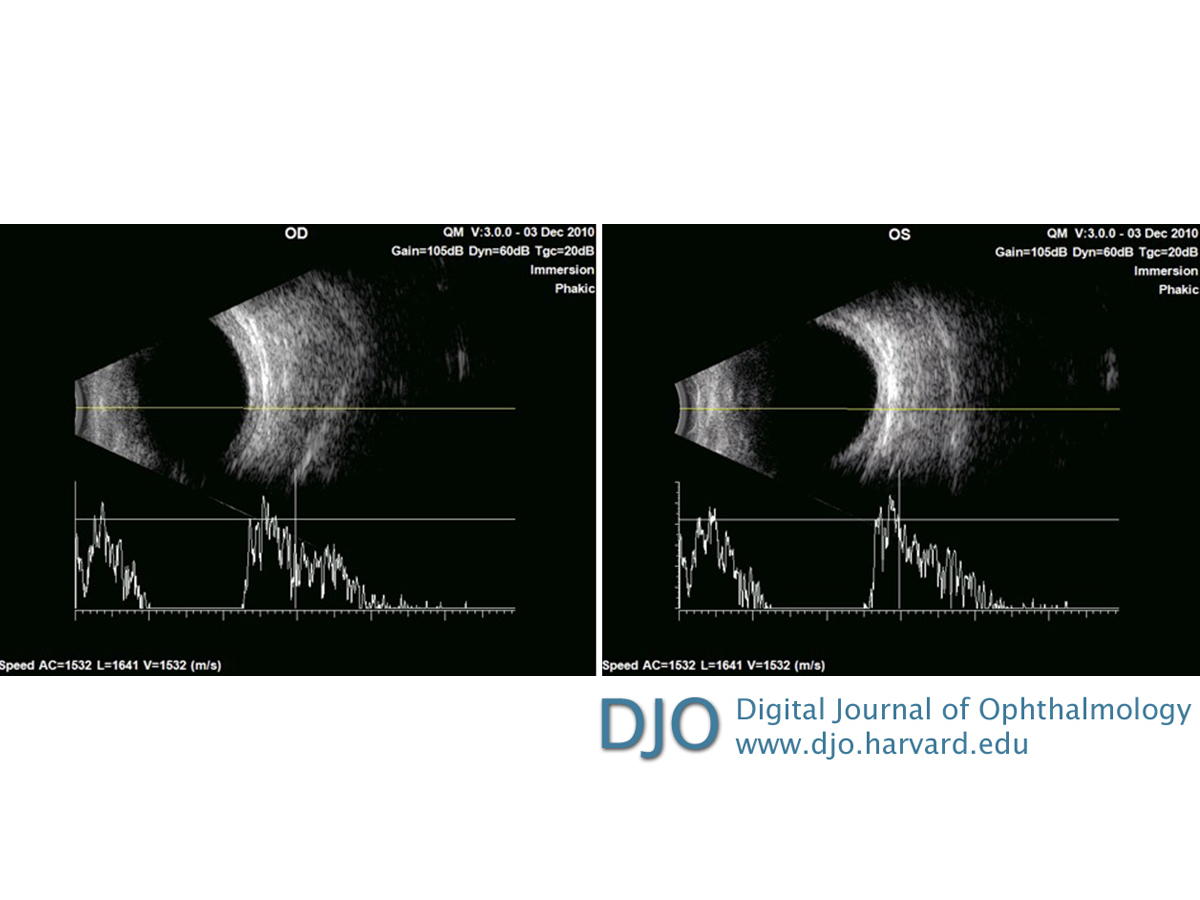
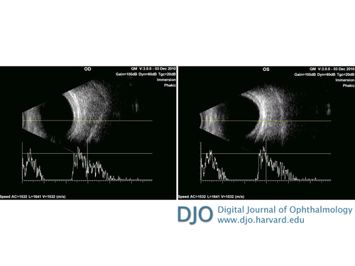
| Ancillary Testing | | Optical coherence tomography showed chorioretinal folds and diffuse intraretinal cystoid changes (Figure 2). In addition to chorioretinal folds, evidence of macular edema was apparent on fluorescein angiography (Figure 3). A-scan ultrasonography revealed an axial length of 19.53 mm in the right eye and 19.48 mm in the left eye. B-scan ultrasonography showed increased retinal-choroidal-scleral thickness in each eye (2.09 mm right eye and 2.36 mm left eye; Figure 4). | |
|
Figure 2
Optical coherence tomography showing choroidal folds and diffuse intraretinal cystoid changes in both eyes, with a central macular thickness of 441 µm in the right eye and 387 µm in the left eye.
 |
|
|
Figure 3
Fluorescein angiography showing alternating hypo- and hyperfluorescent lines in both eyes, corresponding to chorioretinal folds. There was mild early hyperfluorescence in the macula that increased during the course of the study, corresponding to macular edema. Pinpoint hyperfluorescent spots correspond to microaneurysms.
 |
|
|
Figure 4
Ultrasound imaging showing retinal-choroidal-scleral thickness of 2.09 mm in the right eye and 2.36 mm in the left eye. A-scan overlay showing the short axial lengths of each eye, the right eye measuring 19.53 mm and the left eye measuring 19.48 mm.
 |
|
|
|
|
|
|
|
 Welcome, please sign in