|
|
 |
|
|
|
|
|
A 5-year-old girl with decreased vision in the left eye
Digital Journal of Ophthalmology 2015
Volume 21, Number 2
May 9, 2015
DOI: 10.5693/djo.03.2014.09.001
|
Printer Friendly
Download PDF |
|
|

Joel Yap, MBChB | Department of Ophthalmology, Greenlane Clinical Centre, Auckland District Health Board, New Zealand Dianne Sharp, FRANZCO | Department of Ophthalmology, Greenlane Clinical Centre, Auckland District Health Board, New Zealand Shuan Dai, FRANZCO | Department of Ophthalmology, Greenlane Clinical Centre, Auckland District Health Board, New Zealand
|
|
|
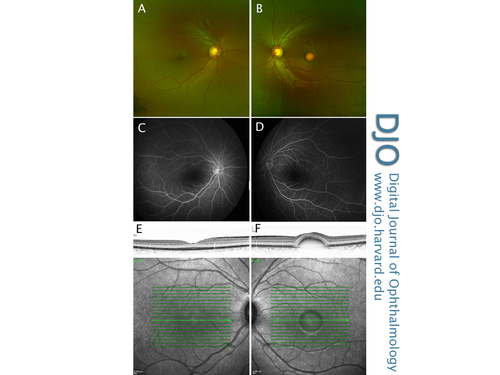
| Examination | | On examination, her best-corrected visual acuity was 20/25 in the right eye and 20/30 in the left eye using a Snellen chart. Frisby stereotest measured stereopsis of 170 arcsec. Ocular motility examination was normal. There was no relative afferent pupillary defect. Anterior segment examination was normal in both eyes; both crystalline lenses were clear. The intraocular pressure was 19 mm Hg in the right eye and 20 mm Hg in the left eye by tonometry (Icare Finland Oy, Helsinki, Finland). Dilated fundus examination revealed a normal right macula and a vitelliform macular lesion in the left eye (Figure 1A,B). The optic nerve heads and peripheral retinae of both eyes appeared otherwise normal. | |
|
Figure 1
Color fundus photographs (A,B), late-stage fundus fluorescein angiogram (C,D) and infrared reflectance imaging with corresponding SD-OCT foveal cross section (E,F) of both the right and left eye, demonstrating asymmetric disease.
 |
|
|
|
|
|
|
|
 Welcome, please sign in


