|
|
 |
|
|
|
|
|
A 31-year-old man with bilateral blurry vision and floaters
Digital Journal of Ophthalmology 2015
Volume 21, Number 2
May 7, 2015
DOI: 10.5693/djo.03.2014.08.003
|
Printer Friendly
Download PDF |
|
|
 Azin Azabari, MD
 | Department of Ophthalmology, Stony Brook University Kevin Kaplowitz, MD | Department of Ophthalmology, Stony Brook University Patrick Sibony, MD | Department of Ophthalmology, Stony Brook University
|
|
|
| Treatment | | The patient was started on 60 mg oral prednisone daily, which was tapered over 1 month. At 1 month after the initial presentation, his visual acuity was 20/20 in each eye. He had mild vitreous cells and mild disc edema bilaterally. In addition, he had developed stellate macular exudates (macular star) in the right eye, retinal vasculitis, and a branch retinal artery and vein occlusion in the left eye (Figure 2). A repeat Bartonella titer was positive at IgG > 1/2560 (negative, <1/320) and IgM 1/200 (negative, <1/100). On further questioning, the patient mentioned he had recently been exposed to a cat. He was started on doxycycline and rifampin. Two months later, his visual acuity remained 20/20, and the disc edema had resolved. The vitritis and macular exudates had cleared completely, but he had developed nasal chorioretinal scars (Figure 3). On his most recent examination, 1 year after initial presentation, his visual acuity was 20/20 in each eye. | |
|
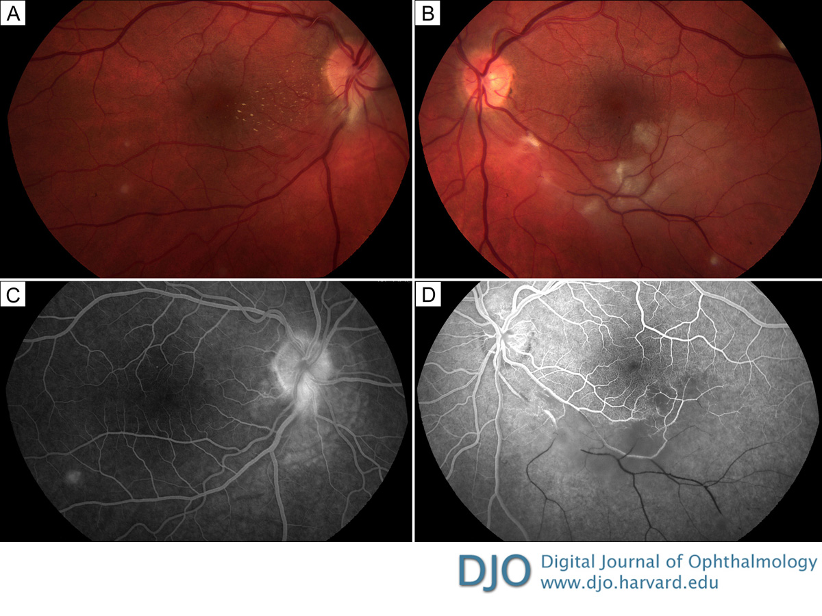
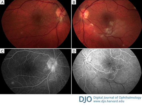
Figure 2
Fundus photographs 1 month after presentation shows stellate macular exudates (macular star) in the right eye (A) as well as a branch retinal artery and vein occlusion in the left eye (B). Representative fluorescein angiography of the right eye (at 1 minute 38 seconds) shows that the inferotemporal site of blockage has cleared (C); in the left eye (at 39 seconds) there is no perfusion distal to the branch retinal artery and vein occlusion with surrounding hypofluorescence from retinal edema present (D).
 |
|
|
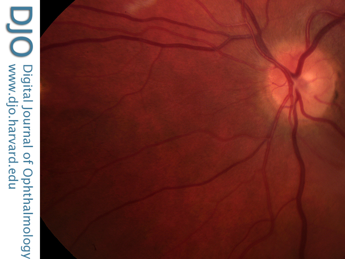
Figure 3
Fundus photograph showing the areas of retinitis becoming chorioretinal scars.
 |
|
|
|
|
|
|
|
 Welcome, please sign in