|
|
 |
|
|
|
|
|
A 31-year-old man with bilateral blurry vision and floaters
Digital Journal of Ophthalmology 2015
Volume 21, Number 2
May 7, 2015
DOI: 10.5693/djo.03.2014.08.003
|
Printer Friendly
Download PDF |
|
|
 Azin Azabari, MD
 | Department of Ophthalmology, Stony Brook University Kevin Kaplowitz, MD | Department of Ophthalmology, Stony Brook University Patrick Sibony, MD | Department of Ophthalmology, Stony Brook University
|
|
|
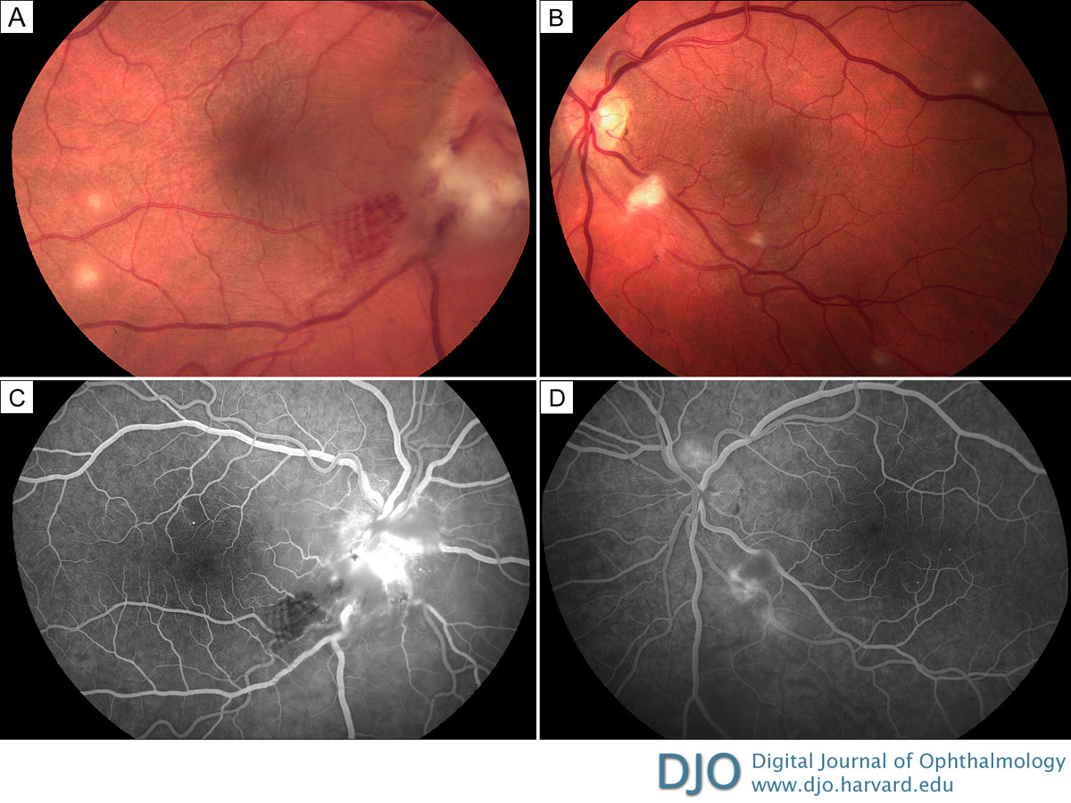
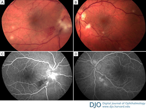
| Examination | | On examination, best-corrected visual acuity was 20/20 in each eye. Slit-lamp examination was remarkable for 1+ anterior chamber cell in both eyes, 3+ anterior vitreous cell in the right eye and +1 in the left eye. Dilated fundus examination disclosed bilateral disc edema, more prominent on the right more than the left, and multifocal deep retinal and choroidal yellowish infiltrates 100–300 μm in diameter (Figure 1). | |
|
Figure 1
Fundus photographs of a 31-year-old man initially presenting with bilateral disc edema, in evidence more on the right (A) than the left (B), and multifocal deep retinal and choroidal yellowish infiltrates 100–300 μm in diameter. Representative fluorescein angiography of the right eye (at 40 seconds) shows prominent optic disc capillary dilation with hyperfluorescence and blockage at the site of the retinitis in the inferotemporal macula (C); the left eye (at 55 seconds) shows prominent optic disc capillary dilation as well as hypofluorescence at the site of the retinitis inferiorly (D).
 |
|
|
|
|
|
|
|
 Welcome, please sign in