|
|
 |
|
|
|
|
|
A 15-year-old girl with variable anisocoria
Digital Journal of Ophthalmology 2014
Volume 20, Number 1
January 12, 2014
DOI: 10.5693/djo.03.2013.10.001
|
Printer Friendly
Download PDF |
|
|

Robert L. Tomsak MD, PhD | Department of Ophthalmology, Kresge Eye Institute, Wayne State University Michael J. Coleman, MD | Department of Ophthalmology, Kresge Eye Institute, Wayne State University
|
|
|
| Examination | On examination, her uncorrected near visual acuity was J1+ and her best-corrected visual acuity was 20/20 at distance and J1+ at near. Her glasses measured −4.50 +0.50 × 98 in the right eye and −4.75 D in the left eye. Ocular motility was full, and the pupils were equal, round, and reactive to light and accommodation. Her pupils measured 5 mm to 3 mm with direct pupillary light reflex and 5 mm to 2.5 mm to near stimulus. Her near point of convergence was approximately 4 cm from the nose, and her accommodative amplitude was assumed normal, given her ease of accommodation and excellent near visual acuity through her full myopic correction. Slit-lamp examination demonstrated normal pupils without iris atrophy, sectoral palsy of the iris sphincter, or vermiform movements.
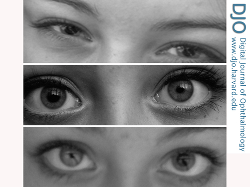
The patient provided several photographs that illustrate fluctuating symptoms. Figure 1 shows inappropriate dilation on a bright sunny day compared to other pictures of her on similar days; there is also subtle anisocoria greater in the right eye than in the left; either pupil could be involved during symptomatic episodes.
| |
|
Figure 1
Photographs demonstrating the variable anisocoria. A, Bilateral mydriasis on a sunny day. B, Right-sided mydriasis. C, Left-sided mydriasis.
 |
|
|
|
|
|
|
|
 Welcome, please sign in


