|
|
 |
|
|
|
|
|
A 50-year-old man with a long-standing, large-angle exotropia and limitation of adduction in the left eye
Digital Journal of Ophthalmology 2013
Volume 19, Number 4
December 30, 2013
DOI: 10.5693/djo.03.2013.09.004
|
Printer Friendly
Download PDF |
|
|

Reshma A. Mehendale, MD | Department of Ophthalmology, Boston Children’s Hospital, and Harvard Medical School, Boston, Massachusetts Anat O. Stemmer-Rachamimov, MD | Department of Pathology, Massachusetts General Hospital, and Ophthalmic Pathology, Massachusetts Eye and Ear Infirmary, and Harvard Medical School Linda R. Dagi, MD | Department of Ophthalmology, Boston Children’s Hospital, and Harvard Medical School, Boston, Massachusetts
|
|
|
| Examination | Best-corrected visual acuity was 20/15 in the right eye and 20/200 in the left eye. There was no afferent pupillary defect, and color vision was full and symmetric. The patient had an exotropia of 40 prism diopters (PD) at distance and 45 PD at near, with a −3 adduction deficit of the left eye greatly increasing the exotropia in right gaze to 60 PD. There was a 2 mm ptosis of the left upper eyelid.
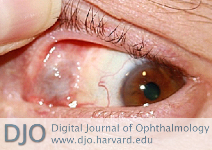
Anterior segment examination revealed conjunctival scarring over the medial and lateral fornices of both eyes. A palpable, painless, soft, purple-gray mass, 8 mm in horizontal diameter and of indeterminate depth was detected in the superonasal aspect of the orbit of the left eye. It extended subconjunctivally posterior to the caruncle and superonasally toward the upper lid. One large superficial conjunctival vessel penetrated the lesion (Figure 1). The bulk of the mass was apparent through closed lids. The patient was unaware of the presence of this mass. The remainder of the ophthalmological examination was unremarkable, including anterior chamber evaluation, intraocular pressure, fundus examination, and visual fields. | |
|
Figure 1
Clinical appearance of a soft, purple-gray subconjunctival mass in the superonasal aspect of the left orbit.
 |
|
|
|
|
|
|
|
 Welcome, please sign in